
Ultrasounds Of The Block
Welcome to SIU's EM Residency "Ultrasounds of the Block" curriculum (formerly known as "Ultrasound of the Month". First instituted as a friendly competition to spark increased volume of bedside studies, it has now become a melee of ultrasound fun. As part of an asynchronous curriculum in emergency ultrasound, all ultrasounds performed at our clinical sites are reviewed for quality assurance by ultrasound director. Each block a few images/clips are selected because of an interesting finding, a high quality image showing great anatomy, or because they carry an important teaching point. These “Ultrasounds of the Block” are then displayed online with clinical vignettes and questions to test residents and their knowledge. The competition aspect of the curriculum is that residents gain points by performing any selected "Ultrasounds of the Block" and for answering monthly questions correctly. Each year a winner is announced based on overall points and surprised with trophy gifts to cherish and gloat about for the many years to come.
Past "Ultrasounds of the Block" can be viewed by clicking here. This blocks "Ultrasounds of the Block" can be viewed below.
Ultrasounds Of The Block (B1)
FAST Learning... and some extra credit...
Question 1:
Left Side |
Right Side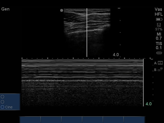
|
The following ultrasound is performed on a trauma patient after they were intubated as part of their airway assessment. Please answer the following questions regarding these images.
1) What is being examined and what probe is being used? (2pts)
2) What ultrasound mode is being used on the left side? (2pts)
3) What ultrasound mode is being used on the right side? (2pts)
4) What is your "sonographic" breathing assessment of this patient? (4pts)
(Courtesy of MK)
1) What is being examined and what probe is being used? (2pts)
2) What ultrasound mode is being used on the left side? (2pts)
3) What ultrasound mode is being used on the right side? (2pts)
4) What is your "sonographic" breathing assessment of this patient? (4pts)
(Courtesy of MK)
Question 2:
After establishing intact airway and breathing, the patient becomes hypotensive. A FAST exam is performed as an adjunct to the trauma patient's circulatory assessment. Please answer the following questions regarding these cardiac images.
|
|
1) What cardiac view is this? (1pts)
2) What is your interpretation? (1pts) (Courtesy of AG & MF) |
|
|
3) What cardiac view is this? (1pts)
4) What is your interpretation? (1pts) (Courtesy of BM) |
|
|
5) What cardiac view is this? (1pts)
6) What is your interpretation? (1pts) 7) What is "X"? (2pts) 8) What is "Y"? (2pts) (Courtesy of MT) |
Question 3:
|
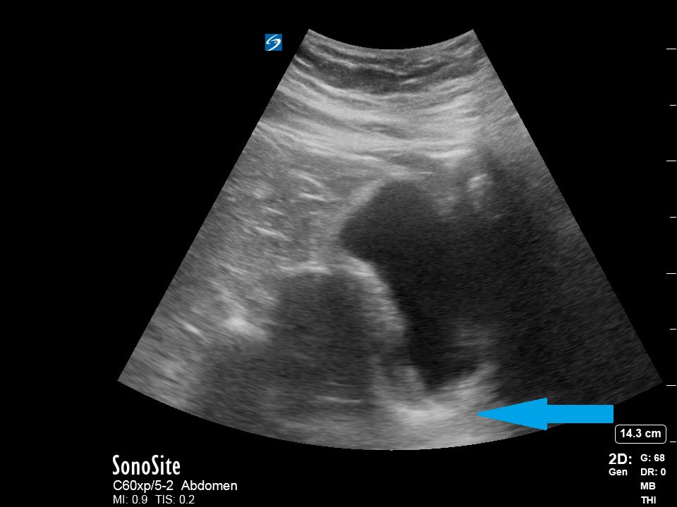
Moving on a RUQ view is obtained on the trauma patient.
1) What probe and mode is being used? (2pts) 2) What three structures should be viewed to ensure an adequate assessment for free fluid in the RUQ view? (2pts) 3) How could this image be optimized? (2pts) 4) What is your interpretation? (4pts) (Courtesy of AG & MF) |
|
Question 4:
|
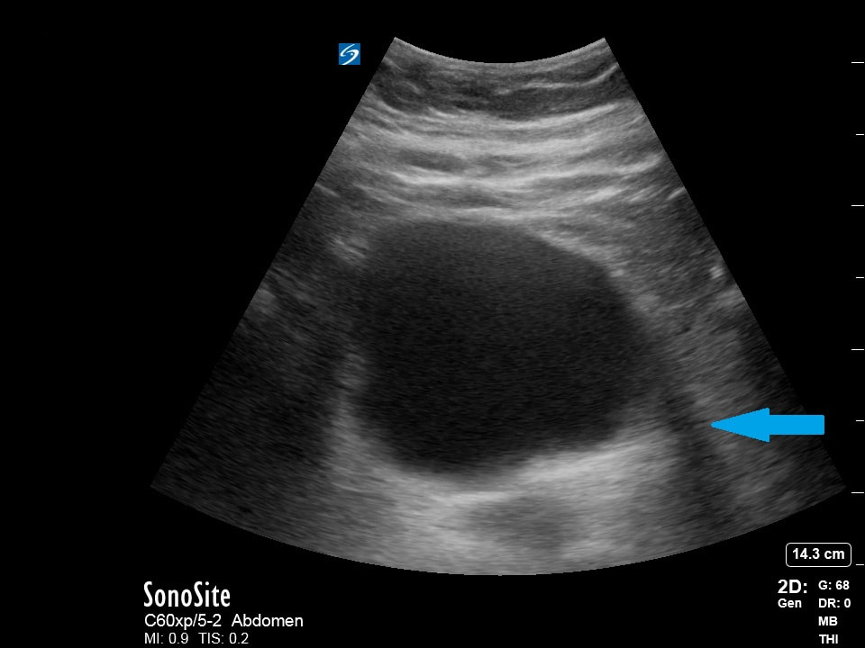
Moving on a LUQ view is obtained on the trauma patient.
1) What probe and mode is being used? (2pts) 2) What three structures should be viewed to ensure an adequate assessment for free fluid in the LUQ view? (2pts) 3) How could this image be optimized? (2pts) 4) What is your interpretation? (4pts) (Courtesy of AG & MF) |
|
Question 5:
Moving on the bladder view is finally assessed on the trauma patient.
|
1) What axis view of the bladder is being assessed? (2pts)
2) What is the name of the artifact seen here? (2pts) (Courtesy of MK) |
|
3) What axis view of the bladder is being assessed? (2pts)
4) What is the name of the artifact seen here? (2pts) (Courtesy of MK) |
|
|
5) What is the name of the artifact seen here? (2pts)
(Courtesy of CP) |
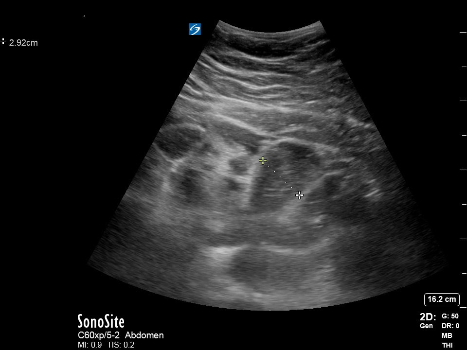
Extra Credit:
|
1) What is the most likely presentation of this patient given this finding? (5pts)
(Courtesy of BK & MW) |
|
|
|
2) What lab result came back positive after hours prompting an urgent care to call this patient and tell them to proceed directly to an emergency department for emergent testing? (5pts)
(Courtesy of CP) |
|
3) What is the most likely presentation of this patient given this finding? (5pts)
(Courtesy of PZ) |
|