
Ultrasounds Of The Block
Welcome to SIU's EM Residency "Ultrasounds of the Block" curriculum (formerly known as "Ultrasound of the Month"). First instituted as a friendly competition to spark increased volume of bedside studies, it has now become a melee of ultrasound fun. As part of an asynchronous curriculum in emergency ultrasound, all ultrasounds performed at our clinical sites are reviewed for quality assurance by ultrasound director. Each block a few images/clips are selected because of an interesting finding, a high quality image showing great anatomy, or because they carry an important teaching point. These “Ultrasounds of the Block” are then displayed online with clinical vignettes and questions to test residents and their knowledge. The competition aspect of the curriculum is that residents gain points by performing any selected "Ultrasounds of the Block" and for answering monthly questions correctly. Each year a winner is announced based on overall points and surprised with trophy gifts to cherish and gloat about for the many years to come.
Past "Ultrasounds of the Block" can be viewed by clicking here. This blocks "Ultrasounds of the Block" can be viewed below.
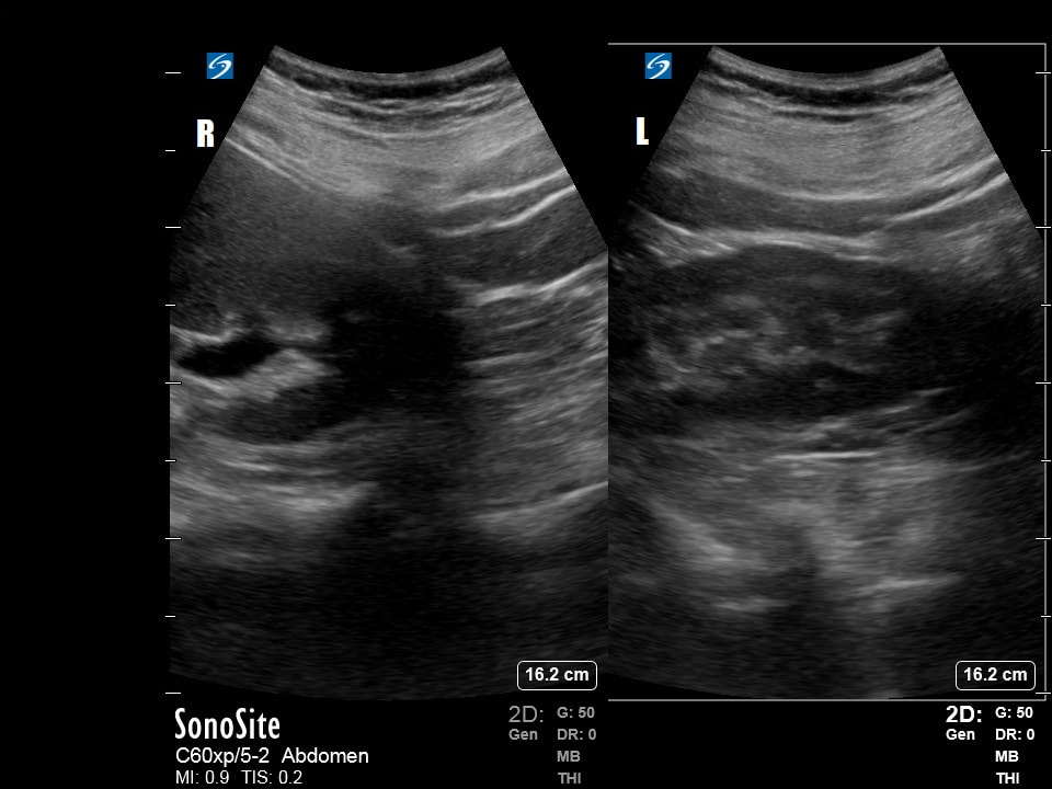
Ultrasounds Of The Block (B4)
Question 1:
Right |
Left |
The following thoracic ultrasound exam is performed on a patient with SOB
a) What probe is being used? (2pts)
b) What are the strengths and limitations of using this probe for this exam? (2pts)
c) What is your interpretation of the images obtained on the right lung? (3pts)
d) What is your interpretation of the images obtained on the left lung? (3pts)
Courtesy of FV & MH
a) What probe is being used? (2pts)
b) What are the strengths and limitations of using this probe for this exam? (2pts)
c) What is your interpretation of the images obtained on the right lung? (3pts)
d) What is your interpretation of the images obtained on the left lung? (3pts)
Courtesy of FV & MH
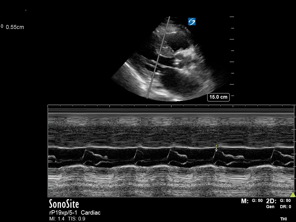
Question 2
|
|
|
The following echo is performed
a) What probe is being used and what view are we in? (2pts)
b) How would you optimize the depth (increase or decrease and why)? (2pts)
c) What is being measured? (2pts)
d) What is your interpretation of the measurement being made and what are you using for your measurement threshold? (4pts)
Courtesy of MH
Question 3:
|
|
The following echo is performed
a) What probe is being used and what view are we in? (2pts) b) How would you optimize the depth (increase or decrease and why)? (2pts) c) What pathology can be visualized? (2pts) d) What anatomical space(s) do you see this pathology? (4pts) Courtesy of FV & MH |
Question 4:
|
|
The following is performed on a patient who passed out at school during PE
a) What probe is being used and what view are we in? (2pts) b) How would you optimize the depth (increase or decrease and why)? (2pts) c) What pathology can be visualized? (2pts) d) What threshold measurement can be used to screen for this condition? (4pts) Courtesy of MM |
Question 5:
|
One of your attending physicians is roomed into one of your rooms in green pod complaining of flank pain. You perform the following ultrasound while he pukes into a green bag infront of his three small children.
a) What probe is being used and at what depth? (2pts) b) What is being examined? (2pts) c) What is your interpretation and most likely diagnosis? (2pts) d) Considering he is scheduled to work in red pod in a couple of hours, the ketorolac you orderd gives him no relief, what is your next analgesic of choice? (4pts) Courtesy of AM |