
Ultrasounds Of The Block
Welcome to SIU's EM Residency "Ultrasounds of the Block" curriculum (formerly known as "Ultrasound of the Month"). First instituted as a friendly competition to spark increased volume of bedside studies, it has now become a melee of ultrasound fun. As part of an asynchronous curriculum in emergency ultrasound, all ultrasounds performed at our clinical sites are reviewed for quality assurance by ultrasound director. Each block a few images/clips are selected because of an interesting finding, a high quality image showing great anatomy, or because they carry an important teaching point. These “Ultrasounds of the Block” are then displayed online with clinical vignettes and questions to test residents and their knowledge. The competition aspect of the curriculum is that residents gain points by performing any selected "Ultrasounds of the Block" and for answering monthly questions correctly. Each year a winner is announced based on overall points and surprised with trophy gifts to cherish and gloat about for the many years to come.
Past "Ultrasounds of the Block" can be viewed by clicking here. This blocks "Ultrasounds of the Block" can be viewed below.
Ultrasounds Of The Block (B5)
Question 1:
|
|
The following ultrasound is obtained on a patient.
a) What is being examined? (2pts) b) What is your interpretation? (2pts) c) Name three different examples of artifact seen on this US? (6pts) Courtesy of AF & EJ |
Question 2:
|
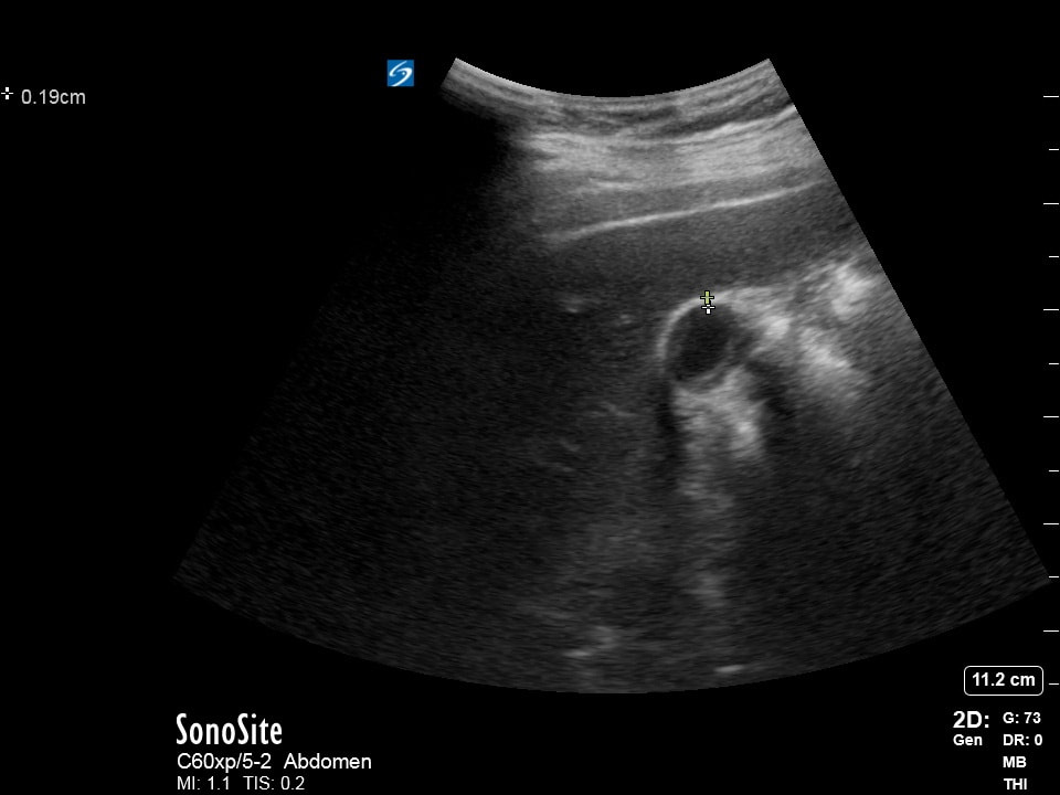
The following ultrasound is obtained.
a) What is being examined? (2pts) b) What is being measured? (2pts) c) What landmark should be included in your image to ensure you are looking at the desired organ? (2pts) d) If the measurement made was actually >0.4cm why would this particular exam be inconclusive? (4pts) Courtesy of ET |
Question 3:
A |
B |
The following ultrasound exam is repeated on the same patient 1 minute apart from each other.
a) What cardiac view is being obtained? (2pts)
b) What landmark are you using to identify your answer in "a"? (2pts)
c) What is your interpretation of the image? (2pts)
d) What did the sonographer change going from exam "A" to exam "B"? (4pts)
Courtesy of ET
a) What cardiac view is being obtained? (2pts)
b) What landmark are you using to identify your answer in "a"? (2pts)
c) What is your interpretation of the image? (2pts)
d) What did the sonographer change going from exam "A" to exam "B"? (4pts)
Courtesy of ET
Question 4:
|
|
The following ultrasound is obtained.
a) What is being examined? (2pts) b) What is your interpretation? (2pts) c) Why did this patient most likely come to the ED? (2pts) d) What is your next step in management for this patient? (4pts) Courtesy of RB |
Question 5:
A |
B |
The following ultrasounds are obtained on spontaneously breathing patients.
a) What is being examined? (2pts)
b) Which patient would you go see first if they presented at the same time? (4pts)
c) If both are hypotensive, which patient(s) might benefit from fluids? (4pts)
Courtesy of BC & DF
a) What is being examined? (2pts)
b) Which patient would you go see first if they presented at the same time? (4pts)
c) If both are hypotensive, which patient(s) might benefit from fluids? (4pts)
Courtesy of BC & DF
Extra Credit:
|
|
Identify the labeled structures (2pts each plus 2pts if all are correct).
a) b) c) d) Courtesy of ZE & AR |