
Ultrasounds Of The Block
Welcome to SIU's EM Residency "Ultrasounds of the Block" curriculum (formerly known as "Ultrasound of the Month"). First instituted as a friendly competition to spark increased volume of bedside studies, it has now become a melee of ultrasound fun. As part of an asynchronous curriculum in emergency ultrasound, all ultrasounds performed at our clinical sites are reviewed for quality assurance by ultrasound director. Each block a few images/clips are selected because of an interesting finding, a high quality image showing great anatomy, or because they carry an important teaching point. These “Ultrasounds of the Block” are then displayed online with clinical vignettes and questions to test residents and their knowledge. The competition aspect of the curriculum is that residents gain points by performing any selected "Ultrasounds of the Block" and for answering monthly questions correctly. Each year a winner is announced based on overall points and surprised with trophy gifts to cherish and gloat about for the many years to come.
Past "Ultrasounds of the Block" can be viewed by clicking here. This blocks "Ultrasounds of the Block" can be viewed below.
Ultrasounds Of The Block (B2)
Question 1:The following FAST Exam was performed on a patient
a) What probe and exam setting was used? (2pts) b) What views are missing? (2pts) c) What is your interpreation? (2pts) d) Provide a clinical vignette that would be consistent with this ultrasound. (4pts + extra for creativity) Courtesy of AF |
|
Question
|
The following ultrasound is performed on a patient who is hypotensive and passed out.
a) What probe and exam setting was used? (2pts) b) What is being examined and in what axis view? (2pts) c) What is your interpretation? (3pts) d) What are your treatment priorities for this patient? (3pts) Courtesy of CY |
|
Question 3:
|
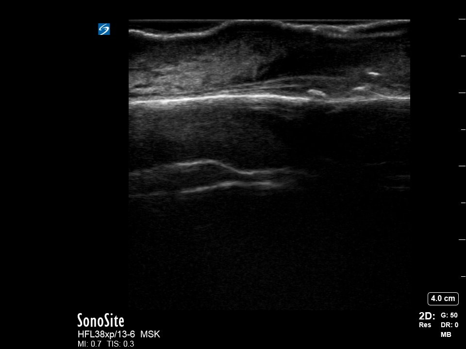
The following ultrasound is performed on a patient with thigh swelling and pain.
a) What probe and exam setting was used? (2pts) b) What is your interpretation? (2pts) c) What "sign" is being tested on this image? (4pts) d) What is your treatment plan for this patient? (2pts) Courtesy of JdC |
|
Question 4:
|
The following ultrasound is performed on a patient a week after suffering and injury to their hand while in an altercation.
a) What probe and exam setting was used? (2pts) b) Besides using gel, what other technique could be used to aquire these images and may improve image quality? (2pts) c) What is your interpretation? (3pts) d) What injury did this patient most likely suffer as a resulf of the altercation? (3pts) Courtesy of RB |
|