
Ultrasounds Of The Block
Welcome to SIU's EM Residency "Ultrasounds of the Block" curriculum (formerly known as "Ultrasound of the Month"). First instituted as a friendly competition to spark increased volume of bedside studies, it has now become a melee of ultrasound fun. As part of an asynchronous curriculum in emergency ultrasound, all ultrasounds performed at our clinical sites are reviewed for quality assurance by ultrasound director. Each block a few images/clips are selected because of an interesting finding, a high quality image showing great anatomy, or because they carry an important teaching point. These “Ultrasounds of the Block” are then displayed online with clinical vignettes and questions to test residents and their knowledge. The competition aspect of the curriculum is that residents gain points by performing any selected "Ultrasounds of the Block" and for answering monthly questions correctly. Each year a winner is announced based on overall points and surprised with trophy gifts to cherish and gloat about for the many years to come.
Past "Ultrasounds of the Block" can be viewed by clicking here. This blocks "Ultrasounds of the Block" can be viewed below.
Ultrasounds Of The Block (B10)
Question 1:
1
|
|
The following PSL echo is obtained on a patient with shortness of breath
a) Traditionally what side of the screen should the apex of the heart be in this view? (2pts) b) What landmark should be included in the bottom of the screen to ensure adequate depth in this view? (2pts) c) What should be done to correct or optimize this view? (2pts) d) What is your interpretation of this ultrasound? (4pts) Courtesy of RB & RL |
Question 2:
|
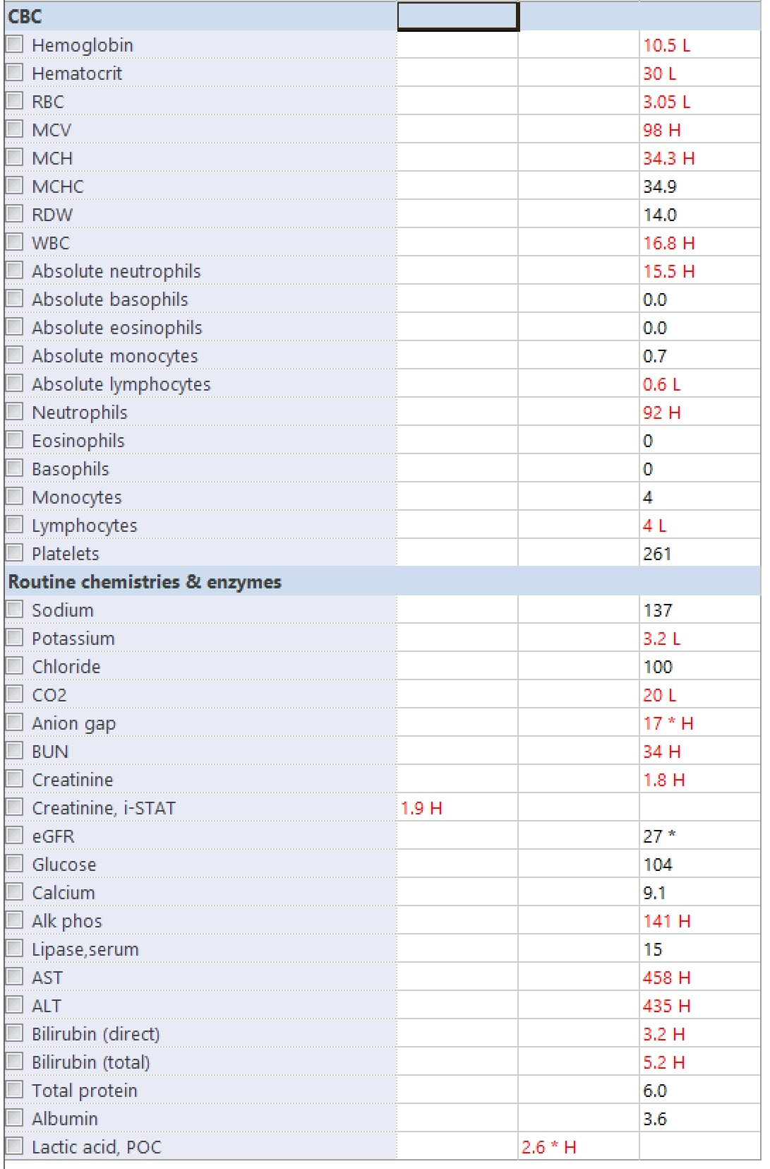
The following labs and ultrasound is obtained on a patient with abdominal pain
a) What landmarks need to be viewed to ensure the gallbladder is being visualized? (2pts) b) What views should be performed to ensure an adequate evaluation of the GB? (2pts) b) Name three findings (in general) on ultrasound that suggest cholecystitis. (6pts) Courtesy of RL |
|
Question 3:
Left |
Right |
The following long axis ultrasound is performed on a patient with knee pain and inability to extend his lower leg.
a) Which knee hurts? (2pts)
b) Is the quadracepts tendon or patellar tendon being evaluated? (4pts)
c) What is your interpretation? (4pts)
Courtesy of DF
a) Which knee hurts? (2pts)
b) Is the quadracepts tendon or patellar tendon being evaluated? (4pts)
c) What is your interpretation? (4pts)
Courtesy of DF
Question 4:
|
|
The following long axis ultrasound is performed on a patient with a positive pregnancy test, vaginal bleeding, and abdominal pain.
a) What could be done to help visualize the uterus better? (3pts) b) What landmarks need to be visualized to ensure the sonographer is viewing the uterus? (3pts) c) What are your next diagnostic steps for this patient? (4pts) Courtesy of RL |
question 5:
|
|
A triage RN asks you to perform a bedside ultrasound of a pregnant patient with abdominal pain. She doesn't know how far along she is and the RN wants you to evaluate if they can go straight to OB triage.
1) What are ACOG's recommendations on what measurements to use on patients in the first trimester? (3pts) 2) What are ACOG's recommendations on what measurements to use on patients in their second and third trimester? (3pts) 3) What trimester and what measurment is the most accurate in dating a pregnancy? (4pts) Courtesy of JB |