
Ultrasounds Of The Block
Welcome to SIU's EM Residency "Ultrasounds of the Block" curriculum (formerly known as "Ultrasound of the Month". First instituted as a friendly competition to spark increased volume of bedside studies, it has now become a melee of ultrasound fun. As part of an asynchronous curriculum in emergency ultrasound, all ultrasounds performed at our clinical sites are reviewed for quality assurance by ultrasound director. Each block a few images/clips are selected because of an interesting finding, a high quality image showing great anatomy, or because they carry an important teaching point. These “Ultrasounds of the Block” are then displayed online with clinical vignettes and questions to test residents and their knowledge. The competition aspect of the curriculum is that residents gain points by performing any selected "Ultrasounds of the Block" and for answering monthly questions correctly. Each year a winner is announced based on overall points and surprised with trophy gifts to cherish and gloat about for the many years to come.
Past "Ultrasounds of the Block" can be viewed by clicking here. This blocks "Ultrasounds of the Block" can be viewed below.
Ultrasounds Of The Block (B9/B10)
Question 1: Too fAST... too Furious
|
|
|
The following images were obtained while performing a FAST exam.
1) What images are missing of this exam? (2 pts)
2) What is wrong with the cardiac view obtained? (4 pts)
3) How would you optimize the images obtained? (4pts)
(Courtesy of DP, MB, RT)
1) What images are missing of this exam? (2 pts)
2) What is wrong with the cardiac view obtained? (4 pts)
3) How would you optimize the images obtained? (4pts)
(Courtesy of DP, MB, RT)
Question 2: Hart's Hearts
|
|
|
The following ultrasound are obtained.
1. What views have been obtained? (2 pts)
2. What could be done to optimize the image quality? (4 pt)
3. What is your interpretation of the ultrasound? (4 pts)
(Courtesy of JH)
1. What views have been obtained? (2 pts)
2. What could be done to optimize the image quality? (4 pt)
3. What is your interpretation of the ultrasound? (4 pts)
(Courtesy of JH)
Question 3: Still Art
1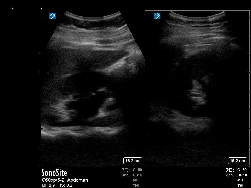
|
2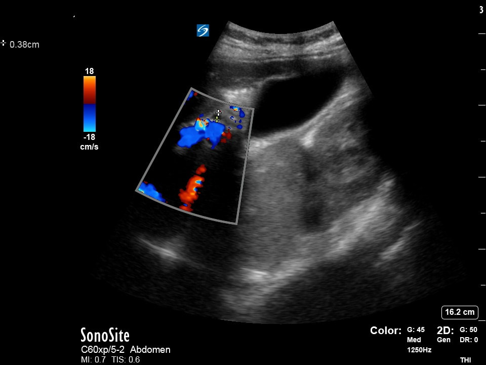
|
3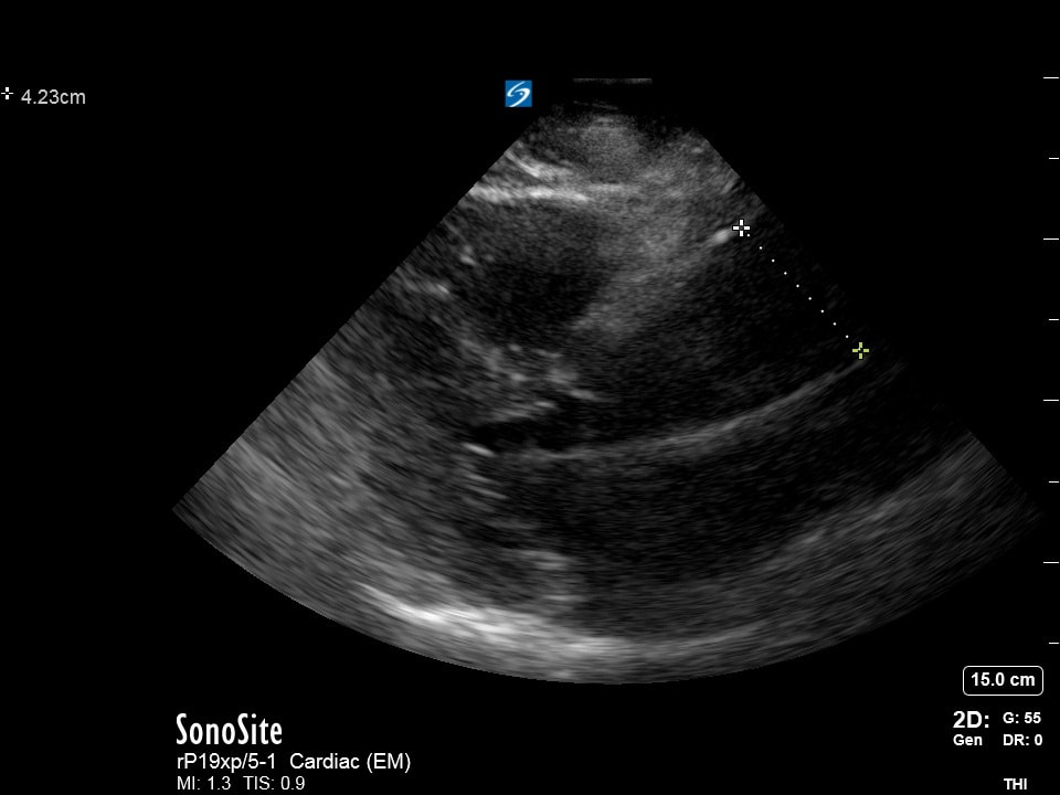
|
State ultrasound mode used, the view obtained, and your interpretation of each still image:
1) (3 pts)
2) (3 pts)
3) (3 pts)
Extra point if you get them all right!
(Courtesy of MB, RT, BK, NF, TF, DP)
1) (3 pts)
2) (3 pts)
3) (3 pts)
Extra point if you get them all right!
(Courtesy of MB, RT, BK, NF, TF, DP)
Question 4: Random access Memory
123 |
State the exam mode, the organ system being examined, and your interpretation:
1) (3 pts) 2) (3 pts) 3) (3 pts) Extra point if you get them all right! (Courtesy of MB, JG, JC) |
Question 5: Dom Dom Dom dom... Dom dom dom dom... dommmmmmmm!!!
|
|
|
A patient presents with diffuse swelling of his hands for months. His joints are swollen and all his fingers are hard to move. The following ultrasound images are obtained of the short axis of his fingers.
1) What could be done to improve the image quality? (2pts)
2) What medium could be used to make this exam easier (i.e. what besides gel could make this a better image?) (4pts)
3) What is your interpretation of the images obtained? (4pts)
(Courtesy of DP)
1) What could be done to improve the image quality? (2pts)
2) What medium could be used to make this exam easier (i.e. what besides gel could make this a better image?) (4pts)
3) What is your interpretation of the images obtained? (4pts)
(Courtesy of DP)
Extra Credit!!!
|
|
|
|
|
The following ultrasounds were obtained on a hypotensive and hypoxic patient.
1) Name all the treatments that may be beneficial for this patient (5pts) 2) Name all the treatments that may be detrimental for this patient (5pts) (Courtesy of AC, ET, RT) |