
Ultrasounds Of The Block
Welcome to SIU's EM Residency "Ultrasounds of the Block" curriculum (formerly known as "Ultrasound of the Month"). First instituted as a friendly competition to spark increased volume of bedside studies, it has now become a melee of ultrasound fun. As part of an asynchronous curriculum in emergency ultrasound, all ultrasounds performed at our clinical sites are reviewed for quality assurance by ultrasound director. Each block a few images/clips are selected because of an interesting finding, a high quality image showing great anatomy, or because they carry an important teaching point. These “Ultrasounds of the Block” are then displayed online with clinical vignettes and questions to test residents and their knowledge. The competition aspect of the curriculum is that residents gain points by performing any selected "Ultrasounds of the Block" and for answering monthly questions correctly. Each year a winner is announced based on overall points and surprised with trophy gifts to cherish and gloat about for the many years to come.
Past "Ultrasounds of the Block" can be viewed by clicking here. This blocks "Ultrasounds of the Block" can be viewed below.
Ultrasounds Of The Block (B6)
Question 1: Heart of my hearts...
|
|
The following ultrasounds are performed on a patient. Please answer the following.
- The probe and exam setting being used (1pts) - The views obtained in order top to bottom (4pts) - List how the image could be optimized (1pts) - Your interpretation of the images (4pts) Extra Credit MCQ (5pts): If the ultrasounds were obtained on a 17 year-old male with recent cough and URI symptoms, what would be the next appropriate test to aid in confirming the diagnosis? (Choose the best answer) A - TEE B - Cardiac Cath C - Myocardial Biopsy D - Cardiac MRI (Courtesy of JB) |
Question 2: Still my beating heart
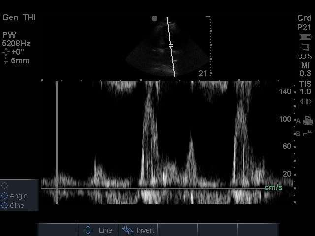
The following ultrasound was performed on a patient with hypotension as part of a calculation to guide fluid resucitation. Please answer the following questions.
- The probe and exam mode setting being used (2pts)
- The cardiac view obtained (2pts)
- The anatomical structure that the calipers are being placed over (3pts)
- The calculation obtained when measuring the "area under the curve" of what is shown (3pts)
- The probe and exam mode setting being used (2pts)
- The cardiac view obtained (2pts)
- The anatomical structure that the calipers are being placed over (3pts)
- The calculation obtained when measuring the "area under the curve" of what is shown (3pts)
Extra Credit MCQ (5pts): What artifact is seen here?
(Choose the best answer)
A - Aliasing
B - Anisotropia
C - Pulse repetition frequency
D - Pulse repetition period
(Courtesy of JH)
(Choose the best answer)
A - Aliasing
B - Anisotropia
C - Pulse repetition frequency
D - Pulse repetition period
(Courtesy of JH)
Question 3: FASTer
|
The following ultrasound if obtained on a patient with transient unilateral extremity weakness. Please list the following:
- The probe and exam setting being used (2pts) - The view obtained (include axis and region/organ being examined) (2pts) - List how the image could be optimized (3pts) - Your interpretation of the image (3pts) |
|
Extra Credit MCQ (5pts): Name the anatomical spaces where fluid is scene on this exam.
(Choose ALL THAT APPLY)
A - Thoracic space
B - Morrison's pouch
C - Pericolic gutter
D - Retrovesicular space
(Courtesy of JC & JB)
(Choose ALL THAT APPLY)
A - Thoracic space
B - Morrison's pouch
C - Pericolic gutter
D - Retrovesicular space
(Courtesy of JC & JB)
Question 4: What am I looking at?
|
|
|
The following ultrasounds are obtained on the same patient.
What medical diagnosis does this patient have? (5pts)
What surgery has this patient had? (5pts)
What medical diagnosis does this patient have? (5pts)
What surgery has this patient had? (5pts)
Extra Credit MCQ (5pts): Please select all that are associated with this patient's diagnosis?
(Choose ALL THAT APPLY)
A - Hypertension
B - SAH
C - Autosomal dominant
D - Autosomal recessive
(Courtesy of JC & ET)
Question 5: Seriously... what am I looking At?
|
|
The following ultrasound if obtained on a patient with transient unilateral extremity weakness. Please list the following:
- The probe and exam setting being used (2pts) - The view obtained (include axis and region/organ being examined) (2pts) - List how the image could be optimized (3pts) - Your interpretation of the image (3pts) (Courtesy of ET) |