
Ultrasounds Of The Block
Welcome to SIU's EM Residency "Ultrasounds of the Block" curriculum (formerly known as "Ultrasound of the Month"). First instituted as a friendly competition to spark increased volume of bedside studies, it has now become a melee of ultrasound fun. As part of an asynchronous curriculum in emergency ultrasound, all ultrasounds performed at our clinical sites are reviewed for quality assurance by ultrasound director. Each block a few images/clips are selected because of an interesting finding, a high quality image showing great anatomy, or because they carry an important teaching point. These “Ultrasounds of the Block” are then displayed online with clinical vignettes and questions to test residents and their knowledge. The competition aspect of the curriculum is that residents gain points by performing any selected "Ultrasounds of the Block" and for answering monthly questions correctly. Each year a winner is announced based on overall points and surprised with trophy gifts to cherish and gloat about for the many years to come.
Past "Ultrasounds of the Block" can be viewed by clicking here. This blocks "Ultrasounds of the Block" can be viewed below.
Ultrasounds Of The Block (B3)
Question 1:The following cardiac ultrasound is performed during a FAST exam
a) What probe and exam setting was used? (2pts) b) How could this image be optimized? (2pts) c) What is anatomical space is this view used to assess for fluid? (2pts) d) Name as many types of artifact that can be seen on this image (1pt for each correct and -1pt for each that is incorrect) Courtesy of MW & AF |
|
Question 2
|
The following RUQ ultrasounds are performed on a patient as the FAST exam is continued on this patient
a) What probe and exam setting was used? (2pts) b) What axis are these images taken in? (2pts) c) What 4 anatomical spaces are you assessing for free fluid in this view? (4pts) d) What artifact do you use in this view to help you evaluate for free fluid not in the abdomen? (2pts) Courtesy of MW & AF |
|
Question 3:
|
The following LUQ ultrasounds are performed on a patient as the FAST exam is continued on this patient
a) What probe and exam setting was used? (2pts) b) What axis are these images taken in? (2pts) c) What 4 anatomical spaces are you assessing for free fluid in this view? (4pts) d) Name the first two spaces where you will most likely see abdominal free fluid in this view? (2pts) Courtesy of MW & AF |
|
Question 4:
|
The following bladder ultrasounds are performed on a patient to complete the FAST exam
a) What probe and exam setting was used? (2pts) b) What axis is this taken in? (2pts) c) Besides looking anterior and posterior to the bladder for free fluid, looking for this increases the sensitivity of this exam for diagnosing interperitoneal injury (2pts) d) Name the 4 most commonly seen artifact that can be seen in this view? (4pts) Courtesy of MW & AF |
|
Question 5
|
The following exam is performed on a patient
a) What probe and exam setting was used? (2pts) b) What exam is being performed? (2pts) c) How would you describe the findings of these images? (4pts) d) What measurement threshold is used here that has a high sensitivity for the diagnosis being targeted here? (2pts) Courtesy of MW & AF 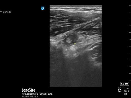
|
|