
Ultrasounds Of The Block
Welcome to SIU's EM Residency "Ultrasounds of the Block" curriculum (formerly known as "Ultrasound of the Month"). First instituted as a friendly competition to spark increased volume of bedside studies, it has now become a melee of ultrasound fun. As part of an asynchronous curriculum in emergency ultrasound, all ultrasounds performed at our clinical sites are reviewed for quality assurance by ultrasound director. Each block a few images/clips are selected because of an interesting finding, a high quality image showing great anatomy, or because they carry an important teaching point. These “Ultrasounds of the Block” are then displayed online with clinical vignettes and questions to test residents and their knowledge. The competition aspect of the curriculum is that residents gain points by performing any selected "Ultrasounds of the Block" and for answering monthly questions correctly. Each year a winner is announced based on overall points and surprised with trophy gifts to cherish and gloat about for the many years to come.
Past "Ultrasounds of the Block" can be viewed by clicking here. This blocks "Ultrasounds of the Block" can be viewed below.
Ultrasounds Of The Block (B3)
Question 1:
|
|
The following ultrasound exam is performed as the first component to a FAST exam.
a) What probe is being used? (2pts) b) What exam mode is being used and what is the benefit of using this exam mode? (2pts) c) What cardiac view is being obtained? (2pts) d) What is your interpretation? (4pts) |
Question 2
|
|
The following ultrasound is performed on a patient as the second component of a FAST exam looking at the RUQ. What are the three anatomical spaces labeled where fluid is seen and for "d" please list the anatomical space that should be assessed in the RUQ that is not visualizeds?
a) (2pts) b) (2pts) c) (2pts) d) (4pts) |
Question 3:
|
|
The follwoing ultrasound is pertormed on a patient as the third component of a FAST exam looking at the LUQ.
a) How could this image be optimized? (2pts) b) What anatomical space is fluid visualized in this image? (2pts) c) What sign is positive in this image that helps confirms where the fluid is? (2pts) d) What artifact allows you you to see this sign? (4pts) |
Question 4:
|
|
The following ultrasound is obtained as the last component of a FAST exam. Name 4 types of artifact seen in this image
a) (2pts) b) (2pts) c) (2pts) d) (4pts) |
Question 5:
|
|
|
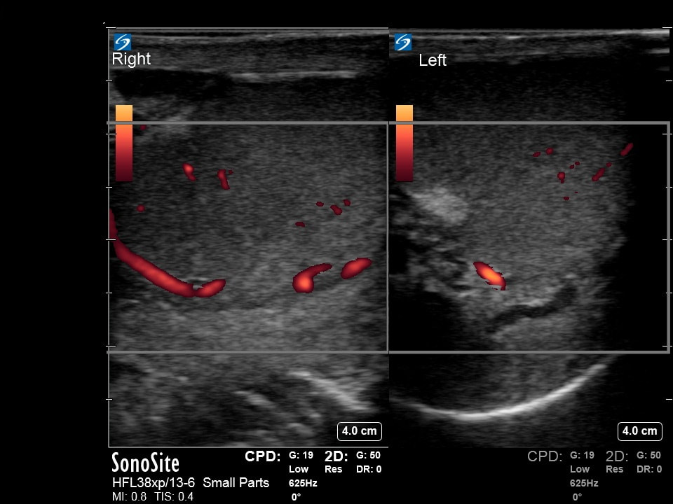
The following ultrasound is obtained on a patient with testicular pain.
a) What probe is being used? (2pts)
b) What does CPD stand for? (2pts)
c) Why is it important to use this mode for this exam? (2pts)
d) What is your interpretation? (4pts)
a) What probe is being used? (2pts)
b) What does CPD stand for? (2pts)
c) Why is it important to use this mode for this exam? (2pts)
d) What is your interpretation? (4pts)
Question 6:
|
|
Provide a clinical vignette for this ultrasound. Points are awarded for correctness of interpretation and creativity. Please note if ChatGPT was used. (10pts)
Courtesy of LN & MH |