
Ultrasounds Of The Block
Welcome to SIU's EM Residency "Ultrasounds of the Block" curriculum (formerly known as "Ultrasound of the Month"). First instituted as a friendly competition to spark increased volume of bedside studies, it has now become a melee of ultrasound fun. As part of an asynchronous curriculum in emergency ultrasound, all ultrasounds performed at our clinical sites are reviewed for quality assurance by ultrasound director. Each block a few images/clips are selected because of an interesting finding, a high quality image showing great anatomy, or because they carry an important teaching point. These “Ultrasounds of the Block” are then displayed online with clinical vignettes and questions to test residents and their knowledge. The competition aspect of the curriculum is that residents gain points by performing any selected "Ultrasounds of the Block" and for answering monthly questions correctly. Each year a winner is announced based on overall points and surprised with trophy gifts to cherish and gloat about for the many years to come.
Past "Ultrasounds of the Block" can be viewed by clicking here. This blocks "Ultrasounds of the Block" can be viewed below.
Ultrasounds Of The Block (B11)
Question 1:
1 |
2 |
1The following US images are obtained while performing a FAST exam on an unstable MVC patient.
a) What views have been included? (2pts)
b) What is your interpretation of image 1? (2pts)
c) What is your interpretation of image 2? (2pts)
d) What lab abnormalities might this patient have? (4pts)
Courtesy of DF
a) What views have been included? (2pts)
b) What is your interpretation of image 1? (2pts)
c) What is your interpretation of image 2? (2pts)
d) What lab abnormalities might this patient have? (4pts)
Courtesy of DF
Question 2:
1 |
2 |
Two separate US images are obtained on two different patients who present after blunt chest trauma.
a) What view has been obtained for both patients? (2pts)
b) Which patient would you be suspicious for a hemothorax? (2pts)
b) What anatomical structure can be visualized in this view when a hemothorax is present and cannot be visualized when a hemothorax is absent? (6pts)
Courtesy of LN
a) What view has been obtained for both patients? (2pts)
b) Which patient would you be suspicious for a hemothorax? (2pts)
b) What anatomical structure can be visualized in this view when a hemothorax is present and cannot be visualized when a hemothorax is absent? (6pts)
Courtesy of LN
Question 3:
The following ultrasound is performed on a patient with vision changes.
a) What is your interpretation/diagnosis? (2pts)
b) What artifact can create the appearance of your suspected diagnosis leading to a false positive interpretation? (4pts)
c) What imaging setting can be turned on to reduce the effect of this artifact? (4pts)
Courtesy of DF
a) What is your interpretation/diagnosis? (2pts)
b) What artifact can create the appearance of your suspected diagnosis leading to a false positive interpretation? (4pts)
c) What imaging setting can be turned on to reduce the effect of this artifact? (4pts)
Courtesy of DF
Question 4:
|
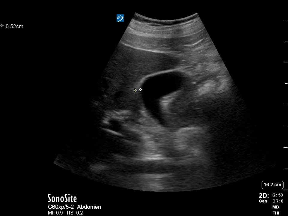
The following ultrasound is obtained while assessing a patient with abdominal pain.
a) What organ is being assessed? (2pts) b) What is being measured? (2pts) c) Why may this measurement lead to a false positive finding? (6pts) |
question 5:
|
|
A patient presents with hip pain. The following ultrasound is performed to assess whether this patient is at risk for a septic joint.
1) Describe the appropriate patient positioning, probe type, and probe placement (including axis) needed to perform this exam? (3pts) 2) What letter corresponds to where a joint effusion needs to be measured? (3pts) 3) What measurement suggests a hip joint effusion? (4pts) Courtesy of JN |