
Ultrasounds Of The Block
Welcome to SIU's EM Residency "Ultrasounds of the Block" curriculum (formerly known as "Ultrasound of the Month"). First instituted as a friendly competition to spark increased volume of bedside studies, it has now become a melee of ultrasound fun. As part of an asynchronous curriculum in emergency ultrasound, all ultrasounds performed at our clinical sites are reviewed for quality assurance by ultrasound director. Each block a few images/clips are selected because of an interesting finding, a high quality image showing great anatomy, or because they carry an important teaching point. These “Ultrasounds of the Block” are then displayed online with clinical vignettes and questions to test residents and their knowledge. The competition aspect of the curriculum is that residents gain points by performing any selected "Ultrasounds of the Block" and for answering monthly questions correctly. Each year a winner is announced based on overall points and surprised with trophy gifts to cherish and gloat about for the many years to come.
Past "Ultrasounds of the Block" can be viewed by clicking here. This blocks "Ultrasounds of the Block" can be viewed below.
Ultrasounds Of The Block (B5)
Question 1: To PE, or not to PE...
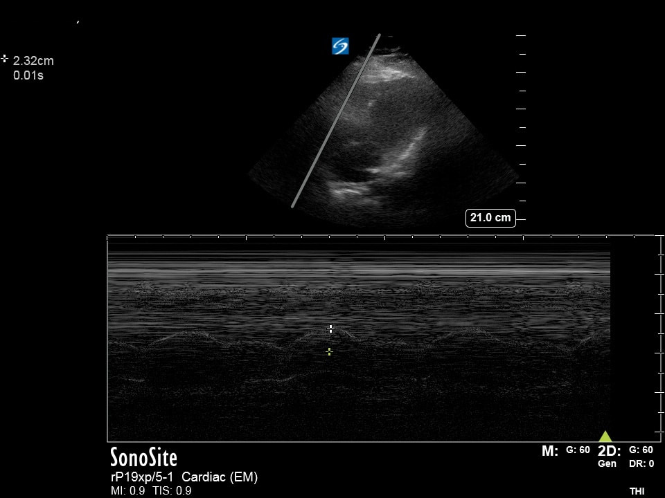
The following ultrasounds are performed on a patient recently discharged from the hospital with a PE. They have been taking their Lovenox and are bridging to coumadin. The were discharged 2 days ago and present to the ED with continued chest pain and stable vitals. Please list:
- The probe and exam setting being used (2pts)
- The view obtained (include axis and region/organ being examined) (2pts)
- List how the image could be optimized (3pts)
- Your interpretation of the image (3pts)
- The probe and exam setting being used (2pts)
- The view obtained (include axis and region/organ being examined) (2pts)
- List how the image could be optimized (3pts)
- Your interpretation of the image (3pts)
A |
B |
|
Extra Credit MCQ (5pts): The following measurement is obtained on the patient which is slightly higher than that measured when discharged. What do you discuss with your patient?
(Choose the best answer) A - You have signs of right heart strain that are worse B - You have signs of right heart strain that are better C - You have signs of right heart strain that are new D - You don't have signs of right heart strain (Courtesy of RT, AC, MM, JB, & DG) |
|
Question 2: A nat o my
The following ultrasound are performed on a patient with flank pain. List the anatomical structures labeled on the exam (A, B, C, D, E) (2pts each).
Extra Credit MCQ (5pts): When performing this exam formally, from what landmarks and in what axis does the aorta need to be visualized to be deemed complete?
(Choose the best answer)
A - Long axis SMA to bifurcation
B - Short axis SMA to bifurcation
C - Long axis celiac trunk to bifurcation
D - Short axis celiac trunk to bifurcation
(Courtesy of DP)
(Choose the best answer)
A - Long axis SMA to bifurcation
B - Short axis SMA to bifurcation
C - Long axis celiac trunk to bifurcation
D - Short axis celiac trunk to bifurcation
(Courtesy of DP)
Question 3: Dopplerganger
Right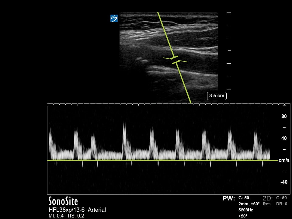
|
Left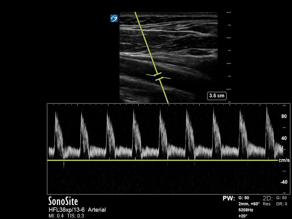
|
The following ultrasound if obtained on a patient with transient unilateral extremity weakness. Please list the following:
- The probe and exam setting being used (2pts)
- The view obtained (include axis and region/organ being examined) (2pts)
- List how the image could be optimized (3pts)
- The side of the body this patient most likely had weakness (3pts)
- The probe and exam setting being used (2pts)
- The view obtained (include axis and region/organ being examined) (2pts)
- List how the image could be optimized (3pts)
- The side of the body this patient most likely had weakness (3pts)
Extra Credit MCQ (5pts): The name of the doppler finding seen here suggesting stenosis in one of the vessels?
(Choose the best answer)
A - Aliasing
B - Anisotropy
C - Spectral broadening
D - Spectral variance
(Courtesy of JC & JB)
(Choose the best answer)
A - Aliasing
B - Anisotropy
C - Spectral broadening
D - Spectral variance
(Courtesy of JC & JB)
Question 4: Finally!!!
|
The following ultrasound if obtained on a patient with a femoral neck fracture. List the anatomical structures labeled (A, B, C, D, E) (2pts each)
|
|
Extra Credit MCQ (5pts): How was this probe placed on the patient?
(Choose the best answer)
A - Long axis (body) medial 1/3 of the inguinal crease
B - Short axis (body) lateral 1/3 of the inguinal crease
C - Short axis (body) middle 1/3 of the inguinal crease
D - Short axis (body) medial 1/3 of the inguinal crease
(Courtesy of RT)
(Choose the best answer)
A - Long axis (body) medial 1/3 of the inguinal crease
B - Short axis (body) lateral 1/3 of the inguinal crease
C - Short axis (body) middle 1/3 of the inguinal crease
D - Short axis (body) medial 1/3 of the inguinal crease
(Courtesy of RT)