
Ultrasounds Of The Block
Welcome to SIU's EM Residency "Ultrasounds of the Block" curriculum (formerly known as "Ultrasound of the Month". First instituted as a friendly competition to spark increased volume of bedside studies, it has now become a melee of ultrasound fun. As part of an asynchronous curriculum in emergency ultrasound, all ultrasounds performed at our clinical sites are reviewed for quality assurance by ultrasound director. Each block a few images/clips are selected because of an interesting finding, a high quality image showing great anatomy, or because they carry an important teaching point. These “Ultrasounds of the Block” are then displayed online with clinical vignettes and questions to test residents and their knowledge. The competition aspect of the curriculum is that residents gain points by performing any selected "Ultrasounds of the Block" and for answering monthly questions correctly. Each year a winner is announced based on overall points and surprised with trophy gifts to cherish and gloat about for the many years to come.
Past "Ultrasounds of the Block" can be viewed by clicking here. This blocks "Ultrasounds of the Block" can be viewed below.
Ultrasounds Of The Block (B6)
Question 1:
|
|
The following ultrasound is performed on a patient during a trauma.
1) What is the technical name for the probe being used and how does it function differently than an abdominal probe? (2pts) 2) What is being exam/view is being obtained? (2pts) 3) What is your interpretation? (2pts) 4) What could be done to optimize the image to see pathology better? (4pts) (Courtesy of BM) |
Question 2:
|
The following ultrasound if performed on an ICU patient
1) What exam/view is being obtained? (2pts) 2) What is your interpretation? (2pts) 3) Describe the maneuvers you would perform on this probe to obtain a perfect exam/view as answered in question "1" ? (2pts) 4) What rotation was Joel probably on when he obtained this and why does it make this image confusing? (4pts) (Courtesy of JG) |
|
Question 3:
|
The following images are obtained on a patient with abdominal pain.
1) What exam/view is being performed? (2pts) 2) What are the two approaches that can be used to perform this exam? (2pts) 3) What landmark needs to be visualized to ensure you are assessing the organ you are looking for on this exam? (2pts) 4) What is your interpretation and what finding (that can be obtained when doing this exam) if positive might prompt a surgical consultation? (4pts) (Courtesy of MK)
|
Question 4:
|
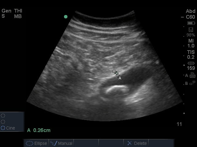
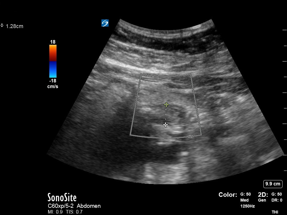
The following ultrasounds are performed. Please list the following regarding each image obtained.
1) What probe is being used? (2 pts) 2) Where has the probe been placed on this patients body? (2 pts) 3) What is your interpretation? (2 pts) 4) What are the parameters that help you make this diagnosis (i.e. measurements)? (4 pts) (Courtesy of MB) |
|
Question 5:
|
|
The following ultrasound is performed on a patient being assessed for abdominal pain and pregnacy.
1) What probe and exam setting was and should be used on this patient? (2pts) 2) What preparations should you make with your patient to optimize your ability to visualize a fetus using transabdominal ultrasound? (2pts) 3) What structures should be visualized and followed to ensure an interuterine pregnancy? (2pts) 4) ALARA calls to utilize ultrasound responsibly using proper settings and scanning times to reduce the risk it poses to the patient. What are the two theoretical risks ultrasound poses to our patients? (4pts) Extra Credit: 1) Given the two theoretical risks ultrasound poses to our patients, each ultrasound machine and its corresponding exam modalities are rated by two indicies. What are they? (4pts) 2) What three physics properties are used to calculate each of these indicies? (6pts) (Courtesy of MK) |