
Ultrasounds Of The Block
Welcome to SIU's EM Residency "Ultrasounds of the Block" curriculum (formerly known as "Ultrasound of the Month"). First instituted as a friendly competition to spark increased volume of bedside studies, it has now become a melee of ultrasound fun. As part of an asynchronous curriculum in emergency ultrasound, all ultrasounds performed at our clinical sites are reviewed for quality assurance by ultrasound director. Each block a few images/clips are selected because of an interesting finding, a high quality image showing great anatomy, or because they carry an important teaching point. These “Ultrasounds of the Block” are then displayed online with clinical vignettes and questions to test residents and their knowledge. The competition aspect of the curriculum is that residents gain points by performing any selected "Ultrasounds of the Block" and for answering monthly questions correctly. Each year a winner is announced based on overall points and surprised with trophy gifts to cherish and gloat about for the many years to come.
Past "Ultrasounds of the Block" can be viewed by clicking here. This blocks "Ultrasounds of the Block" can be viewed below.
Ultrasounds Of The Block (B4)
Question 1: Tom Sawyer
The following ultrasounds are performed on 2 patients complaining of difficulty in breathing. Please list the following for each patient.
- The probe and exam setting being used (2pts)
- The view obtained (include axis and region/organ being examined) (2pts)
- List how the image could be optimized (3pts)
- Your interpretation of the image (3pts)
- The probe and exam setting being used (2pts)
- The view obtained (include axis and region/organ being examined) (2pts)
- List how the image could be optimized (3pts)
- Your interpretation of the image (3pts)
Right |
Left |
|
|
|
Extra Credit MCQ (5pts): When examining this patient what would you expect to hear on their lung exam?
(Choose the best answer)
A - Clear lung sounds b/l
B - Absent lung sounds on the right
C - Crackles on the left
D - Crackles on the right
(Courtesy of MW)
(Choose the best answer)
A - Clear lung sounds b/l
B - Absent lung sounds on the right
C - Crackles on the left
D - Crackles on the right
(Courtesy of MW)
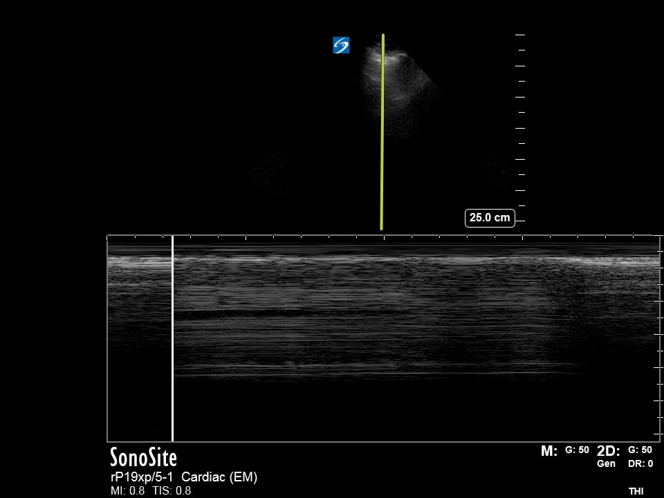
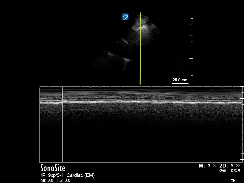
Question 2: The spirit of radio
The following ultrasounds are performed on 2 patients complaining of difficulty in breathing. Please list the following for each patient.
- The probe and exam setting being used (2pts)
- The view obtained (include axis and region/organ being examined) (2pts)
- List how the image could be optimized (3pts)
- Your interpretation of the image (3pts)
- The probe and exam setting being used (2pts)
- The view obtained (include axis and region/organ being examined) (2pts)
- List how the image could be optimized (3pts)
- Your interpretation of the image (3pts)
Right |
Left |
|
|
Extra Credit MCQ (5pts): The patient suddenly becomes tachy and hypotensive. What is the next best course of action?
(Choose the best answer)
A - Needle decompression on the Left
B - Tube thoracostomy on the left
C - Needle decompression on the right
D - Tube thoracostomy on the right
(Courtesy of DF)
(Choose the best answer)
A - Needle decompression on the Left
B - Tube thoracostomy on the left
C - Needle decompression on the right
D - Tube thoracostomy on the right
(Courtesy of DF)
Question 3: Fly by night
|
|
The following ultrasound if obtained on a hypotensive patient in the ICU. Please answer the following for each image:
- The probe and exam setting being used (2pts) - The view obtained (include axis and region/organ being examined) (2pts) - List how the image could be optimized (3pts) - Your interpretation of the image (3pts) |
Extra Credit MCQ (5pts): What is a key piece of information needed to appropriately interpret this image to guide resuscitation efforts?
(Choose the best answer)
A - Whether or not the patient has crackles on lung exam
B - Whether or not the patient has edema in their extremities
C - Whether or not the patient is being assisted with ventillation
D - Wheather or not the patient has JVD
(Courtesy of JC)
(Choose the best answer)
A - Whether or not the patient has crackles on lung exam
B - Whether or not the patient has edema in their extremities
C - Whether or not the patient is being assisted with ventillation
D - Wheather or not the patient has JVD
(Courtesy of JC)
Question 4: Limelight
|
|
The following ultrasound if obtained on a hypotensive patient in the ED. Please answer the following for each image:
- The probe and exam setting being used (2pts) - The view obtained (include axis and region/organ being examined) (2pts) - List how the image could be optimized (3pts) - Your interpretation of the image (3pts) |
Extra Credit MCQ (5pts): The patient is pale, tachycardic, tachypeneic, and hypotensive. What is the most appropriate next step in management?
(Choose the best answer)
A - Endotracheal intubation
B - Intravenous crystalloids
C - tPA IV
D - Inotropic agent IV
(Courtesy of ET)
(Choose the best answer)
A - Endotracheal intubation
B - Intravenous crystalloids
C - tPA IV
D - Inotropic agent IV
(Courtesy of ET)
Extra Credit (Q1-Q4)
Extra Credit (5pts): What is the connection of the question titles #1-4 and the images presented?
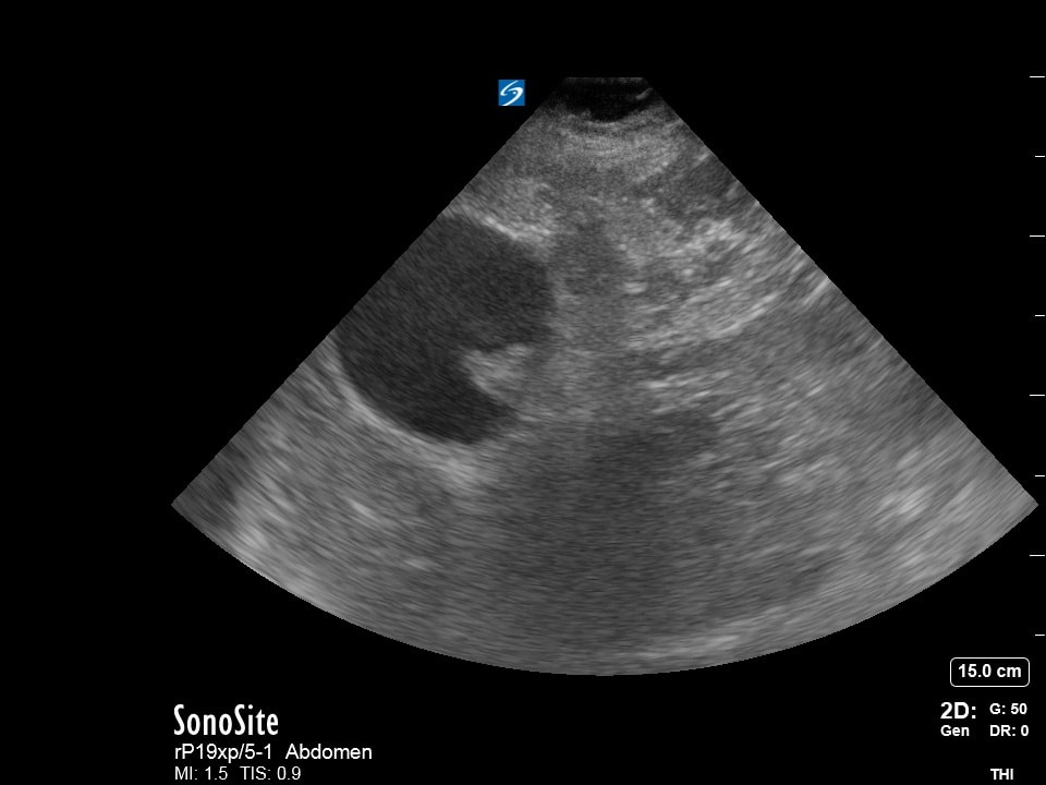
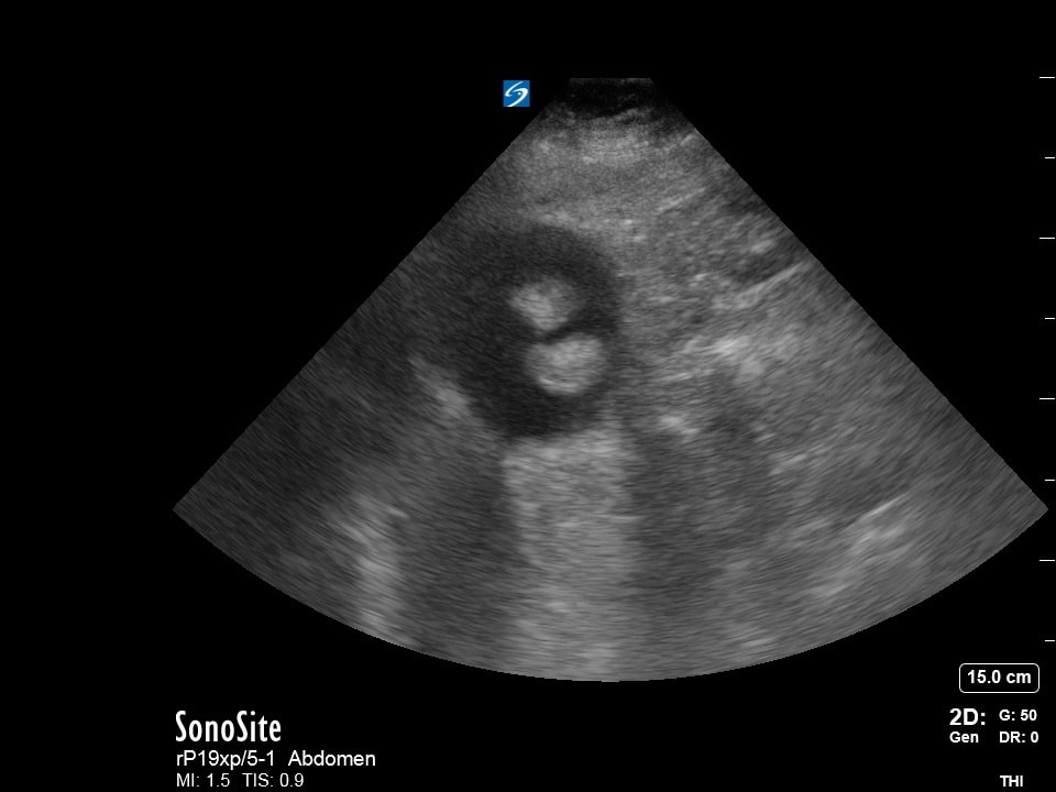
Question 5: Gallblassen Eins
|
The following ultrasound is obtained on a patient with abdominal pain. Provide the following.
The probe and exam setting being used (2pts) The view obtained (include axis and region/organ being examined) (2pts) List how the image could be optimized (3pts) Your interpretation of the image (3pts) (Courtesy of JG) |
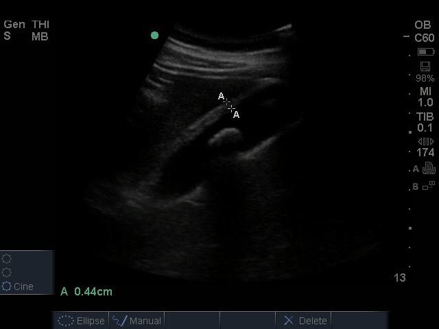
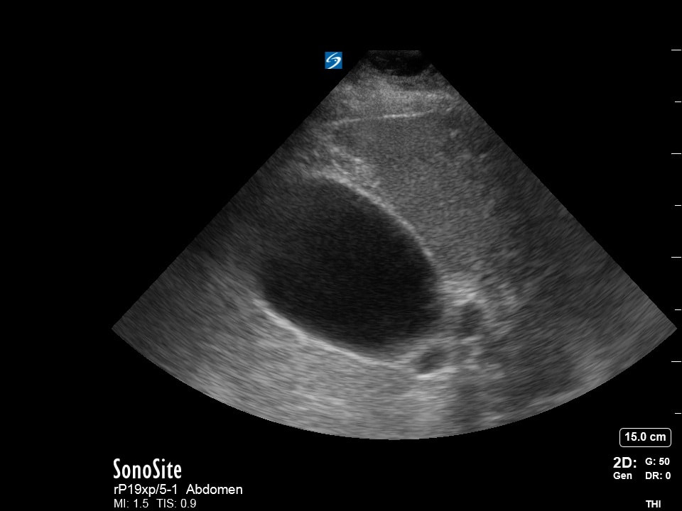
Question 6: Gallblassen Zwei
|
|
|
The following ultrasound is obtained on a patient with abdominal pain. Provide the following.
The probe and exam setting being used (2pts)
The view obtained (include axis and region/organ being examined) (2pts)
List how the image could be optimized (3pts)
Your interpretation of the image (3pts)
(Courtesy of DP)
The probe and exam setting being used (2pts)
The view obtained (include axis and region/organ being examined) (2pts)
List how the image could be optimized (3pts)
Your interpretation of the image (3pts)
(Courtesy of DP)