
Ultrasounds Of The Block
Welcome to SIU's EM Residency "Ultrasounds of the Block" curriculum (formerly known as "Ultrasound of the Month". First instituted as a friendly competition to spark increased volume of bedside studies, it has now become a melee of ultrasound fun. As part of an asynchronous curriculum in emergency ultrasound, all ultrasounds performed at our clinical sites are reviewed for quality assurance by ultrasound director. Each block a few images/clips are selected because of an interesting finding, a high quality image showing great anatomy, or because they carry an important teaching point. These “Ultrasounds of the Block” are then displayed online with clinical vignettes and questions to test residents and their knowledge. The competition aspect of the curriculum is that residents gain points by performing any selected "Ultrasounds of the Block" and for answering monthly questions correctly. Each year a winner is announced based on overall points and surprised with trophy gifts to cherish and gloat about for the many years to come.
Past "Ultrasounds of the Block" can be viewed by clicking here. This blocks "Ultrasounds of the Block" can be viewed below.
Ultrasounds Of The Block (B2)
From head to Groin... and all the area in between
Question 1:
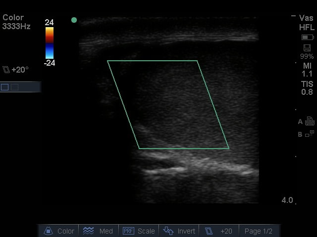
Clip 1
|
Clip 2
|
The following ultrasounds are performed on a hypotensive patient as part of the RUSH exam. Please answer the following questions regarding these images.
1) What is being examined and what probe is being used? (2pts)
2) What are the anechoicc structures seen transversing the screen horizontally in clip 1 and which one is visualized best in clip 2? (2pts)
Hint: Use both videos to get a nice view of one of the structures.
3) How do you use the IVC to assess fluid status? Include the landmarks for measurements in your answer. (2pts)
4) How does the probe need to be manipulated to fully visualize structure A in clip 1? (4pts)
(Courtesy of CP)
Question 2:
A 34 year old male presents to the ER with left flank pain and hematuria. After fluids and providing a urine sample, he is feeling much better. Please answer the following questions regarding this clip.
|
|
1) What is being evaluated? (2pts)
2) What mode is being used? (2pts) 3) What is your interpretation of the clip? (3pts) 4) What is your diagnosis and management? (3pts) (Courtesy of MB) |
Question 3:
|
A severely near sighted patient comes to the ER after falling down some stairs at the grandstand at the state fair. There are bruises all over the face from where it hit the bench below.
1) What exam is being performed? (2pts) 2) What is your interpretation? (2pts) (Courtesy of BK & JG) |
|
The patient was admitted to the hospital due to inability to clear the C collar while under the influence. After going through the DTs, he started complaining of some worsening leg pain so the following images were obtained.
3) What is your interpretation? (2pts)
4) What is the management? (4pts)
(Courtesy of MB)
3) What is your interpretation? (2pts)
4) What is the management? (4pts)
(Courtesy of MB)
Left Leg
|
Right Leg
|
Question 4:
|
Phil has been living a pretty safe life while here in Springfield. When Jaylen learned to talk, his favorite thing to say was Ball! He threw a fastball at dad and dad started having some pain. He was a bit nervous, so he called on his fellow dad Caleb to lend him a hand and see if they could figure out if he was ok.
1) What is being examined? (2 points) 2) What mode is being used? (2 points) 3) What is your interpretation as to the cause of Phil's pain? (2 points) (Courtesy of CP) After being terrified by the dangers of life on the farm, he ditched the "Bullpen" briefs and upgraded to his Minnesota and deer hunting roots with the new Duluth Trading Company "Buck Naked Underwear." Then he started having some pain. Caleb had already had enough of Phil's schenanigans so he decided to call on Tyler this time.
4) What is your interpretation and disposition? (4pts) (Courtesy of TF) |
|
Question 5:
A series of patients all come into the emergency department with chest pain and shortness of breath. Rapid diagnoses and dispositions were made by looking at the chest instead of just trying to listen to it or percuss it.
Patient 1: Mr. McConnell
|
|
1) What probe and exam mode are being used? (1pt)
2) Which view is this? (1pt) 3) What is your interpretation? (2pts) (Courtesy of Marler Feet) |
Patient 2: Mr. Watson
|
|
|
4) Where is this probe placed and how is it oriented? (2pts)
5) What is the anechoic circle below the diaphragm in the first clip? (2pts)
6) What is your interpretation for this patient based on both clips above? (2pts)
(Courtesy of MT & NF)
5) What is the anechoic circle below the diaphragm in the first clip? (2pts)
6) What is your interpretation for this patient based on both clips above? (2pts)
(Courtesy of MT & NF)
Extra Credit:
|
A 44 YOF presents to the ER after eating a horseshoe at Obed's.
1) What is the differential diagnosis given the findings? (2pts) 2) What is your interpretation of the image? (3 pts) (Courtesy of CP) |
|
This patient presented as a Cat I trauma after a high speed MVC.
3) Tell the narrative of what is happening for this patient based on this series of clips. (5 pts)
(Courtesy of MK)
3) Tell the narrative of what is happening for this patient based on this series of clips. (5 pts)
(Courtesy of MK)
Clip 1
|
Clip 2 |
|
|
|
Clip 3 |
Clip 4 |
|
|
|