
Ultrasounds Of The Block
Welcome to SIU's EM Residency "Ultrasounds of the Block" curriculum (formerly known as "Ultrasound of the Month"). First instituted as a friendly competition to spark increased volume of bedside studies, it has now become a melee of ultrasound fun. As part of an asynchronous curriculum in emergency ultrasound, all ultrasounds performed at our clinical sites are reviewed for quality assurance by ultrasound director. Each block a few images/clips are selected because of an interesting finding, a high quality image showing great anatomy, or because they carry an important teaching point. These “Ultrasounds of the Block” are then displayed online with clinical vignettes and questions to test residents and their knowledge. The competition aspect of the curriculum is that residents gain points by performing any selected "Ultrasounds of the Block" and for answering monthly questions correctly. Each year a winner is announced based on overall points and surprised with trophy gifts to cherish and gloat about for the many years to come.
Past "Ultrasounds of the Block" can be viewed by clicking here. This blocks "Ultrasounds of the Block" can be viewed below.
Ultrasounds Of The Block (B7)
Question 1:
Left |
Right |
The following ultrasound exam is performed on a patient with flank pain
a) What probe and exam setting is used? (2pts)
b) What is the depth? (2pts)
c) What axis are these ultrasounds taken in relelation to the body? (2pts)
d) What side is this patient having flank pain? (2pts)
e) In what patient populations would performing this ultrasound be sufficient in diagnosing ureteral lithiasis without any other imaging? (2pts)
Courtesy of KK
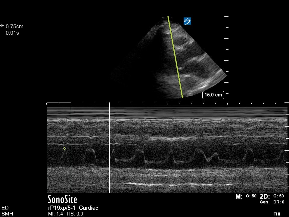
Question 2
|
The following ultrasound is obtained on a patient
a) What probe and exam mode was used? (2pts) b) What measurement is being obtained? (2pts) c) What cardiac view is used to obtain this measurement? (2pts) d) What is the threshold used to identify abnormality with this measurement? (4pts) Courtesy of KK |
|
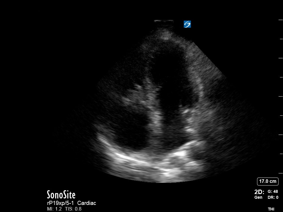
Question 3:
|
The following echo is performed on a hypoxic patient
a) What cardiac view is being obtained? (2pts) b) What could be done to optimize this image? (2pts) c) What sign is seen here and how would you describe it? (2pts) d) What is this sign highly specific for? (4pts) Courtesy of Dan-Don |
|
Question 4:
|
|
|
The following echo is performed on a patient who later left AMA
a) What cardiac view was obtained in the video image? (2pts)
b) What cardiac view was obtained on the still image? (2pts)
c) What is your interpretation? (2pts)
d) Provide a clinical vignette for this encounter? (4pts)
Courtesy of BC
a) What cardiac view was obtained in the video image? (2pts)
b) What cardiac view was obtained on the still image? (2pts)
c) What is your interpretation? (2pts)
d) Provide a clinical vignette for this encounter? (4pts)
Courtesy of BC