
Ultrasounds Of The Block
Welcome to SIU's EM Residency "Ultrasounds of the Block" curriculum (formerly known as "Ultrasound of the Month". First instituted as a friendly competition to spark increased volume of bedside studies, it has now become a melee of ultrasound fun. As part of an asynchronous curriculum in emergency ultrasound, all ultrasounds performed at our clinical sites are reviewed for quality assurance by ultrasound director. Each block a few images/clips are selected because of an interesting finding, a high quality image showing great anatomy, or because they carry an important teaching point. These “Ultrasounds of the Block” are then displayed online with clinical vignettes and questions to test residents and their knowledge. The competition aspect of the curriculum is that residents gain points by performing any selected "Ultrasounds of the Block" and for answering monthly questions correctly. Each year a winner is announced based on overall points and surprised with trophy gifts to cherish and gloat about for the many years to come.
Past "Ultrasounds of the Block" can be viewed by clicking here. This blocks "Ultrasounds of the Block" can be viewed below.
Ultrasounds Of The Block (B5)
Question 1:
|
|
The following ultrasound is performed on a patient during a trauma.
1) Is this a LUQ or RUQ view? (2pts) 2) What is your reasoning for what view it is? (2pts) 3) What is your interpretation? (2pts) 4) What could be done to optimize the image to see pathology better? (4pts) (Courtesy of CP) |
Question 2:
|
A patient presents with sudden vision loss.
1) What probe is being used? (2pts) 2) What is being examined? (2pts) 3) What is your interpreation? (2pts) 4) What could be done to optimize this image to see pathology better? (4pts) (Courtesy of CP) |
|
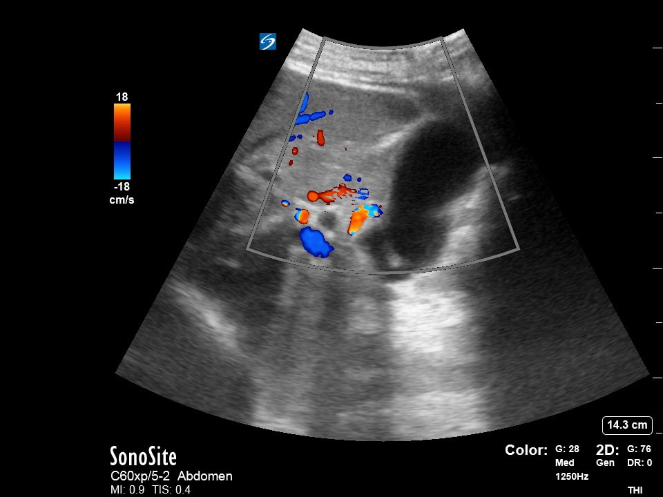
Question 3:
|
|
The following images are obtained on a patient with flank pain.
1) What is your interpretation of the bladder view? (2pts) 2) What is your interpretation of Clip A? (2pts) 3) What is your interpretation of Clip B? (2pts) 4) Assuming James used traditional probe orientation to obtain his short axis view of the bladder, which side is Clip A and which side is Clip B? (4pts) |
(Courtesy of JH)
Clip A(Courtesy of MB)
|
Clip B(Courtesy of CP)
|
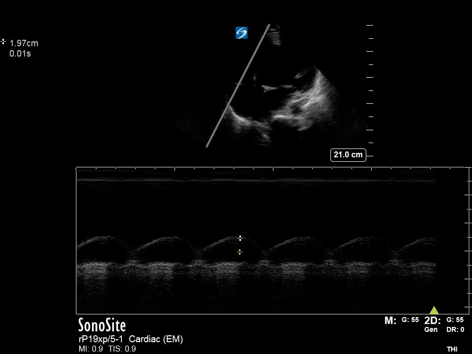
Question 4:
|
The following cardiac ultrasounds are performed. Please list the following regarding each image obtained.
1) What view and what findings? (2 pts) 2) What view and what findings? (2 pts) 3) What measurement is being obtained and your interpretation? (3 pts) 4) What measurement is being obtained and your interpretation? (3 pts) (Courtesy of ET, JG, JC, AC, & MB) |
1)2)3)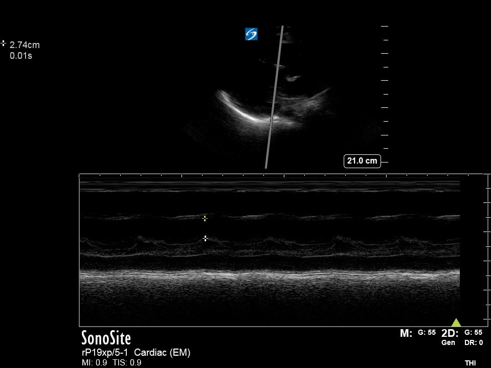
4)
|
Question 5:
|
|
|
Please answer the following (Make sure your audio is working!!!).
1) What is "A"? (2pts)
2) What is "B"? (2pts)
3) What is "C"? (2pts)
4) What is "D"? (4pts)
Extra Credit: Who was called upon to help us out with interpreting this ultrasound? (5pts!!!)
(Courtesy of KG & NF)
1) What is "A"? (2pts)
2) What is "B"? (2pts)
3) What is "C"? (2pts)
4) What is "D"? (4pts)
Extra Credit: Who was called upon to help us out with interpreting this ultrasound? (5pts!!!)
(Courtesy of KG & NF)
Extra Credit:
|
The following ultrasound is performed.
1) What is "A"? (2pts) 2) What is "B"? (2pts) 3) What is "C"? (2pts) 4) Why is knowing this anatomy important? (4pts) (Courtesy of BM, JH, & ET) |
|