
Ultrasounds Of The Block
Welcome to SIU's EM Residency "Ultrasounds of the Block" curriculum (formerly known as "Ultrasound of the Month"). First instituted as a friendly competition to spark increased volume of bedside studies, it has now become a melee of ultrasound fun. As part of an asynchronous curriculum in emergency ultrasound, all ultrasounds performed at our clinical sites are reviewed for quality assurance by ultrasound director. Each block a few images/clips are selected because of an interesting finding, a high quality image showing great anatomy, or because they carry an important teaching point. These “Ultrasounds of the Block” are then displayed online with clinical vignettes and questions to test residents and their knowledge. The competition aspect of the curriculum is that residents gain points by performing any selected "Ultrasounds of the Block" and for answering monthly questions correctly. Each year a winner is announced based on overall points and surprised with trophy gifts to cherish and gloat about for the many years to come.
Past "Ultrasounds of the Block" can be viewed by clicking here. This blocks "Ultrasounds of the Block" can be viewed below.
Ultrasounds Of The Block (B9)
Question 1:
|
|
|
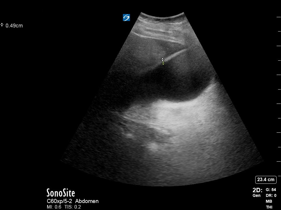
A patient who is presents with abdominal pain after evaluated by a provider in an upfront process. Their vitals are unremarkable and a comprehensive metabolic panel is ordered and without abnormalities. The following ultrasound is ordered as part of an assessment for gallbladder pathology. What exam finding would help aid in interpreting the images and measurement obtained? (10pts)
a) diffuse abdominal tenderness to palpation
b) caput medusae
c) pale tongue and inner eyelids
d) pulsating abdominal mass
Courtesy of MM
a) diffuse abdominal tenderness to palpation
b) caput medusae
c) pale tongue and inner eyelids
d) pulsating abdominal mass
Courtesy of MM
Question 2
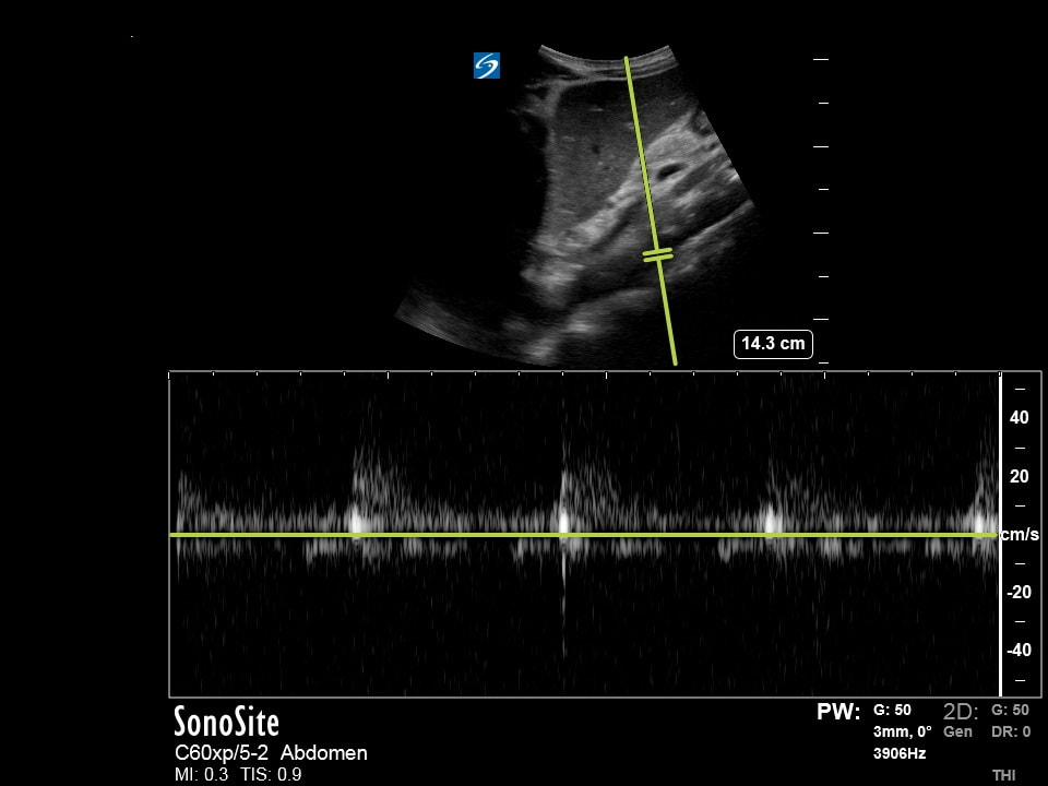
With regards to the exam peformed in "Question 1", what other situations would make interpreting the images and measurements obtained difficult? (10pts)
a) history of peritoneal dialysis
b) obtained postprandial
c) obtained fasting
d) "a" and "b"
e) "a" and "c"
Courtesy of MM
a) history of peritoneal dialysis
b) obtained postprandial
c) obtained fasting
d) "a" and "b"
e) "a" and "c"
Courtesy of MM
Question 3:
The following ultrasound is performed on a patient with abdominal pain and vomiting. What constellation of findings is suggestive of small bowel obstruction for this patient? (10pts)
a) fluid filled bowel, > 3cm diameter in caliber, with peristalsis
b) air filled bowel, > 3cm diameter in caliber, without peristalsis
c) fluid filled bowel, > 2cm diameter in caliber, without peristalsis
d) air filled bowel, > 2cm diameter in caliber, with peristalsis
Courtesy of MM
a) fluid filled bowel, > 3cm diameter in caliber, with peristalsis
b) air filled bowel, > 3cm diameter in caliber, without peristalsis
c) fluid filled bowel, > 2cm diameter in caliber, without peristalsis
d) air filled bowel, > 2cm diameter in caliber, with peristalsis
Courtesy of MM
Question 4:
The following images is obtained during a RUSH exam. Assuming the sonographer was correctly identifying a specific structure of interest, what were they assessing for in this view? (10pts)
a) assessing for obstructive shock
b) identifying a source of obstructive shock
c) assessing for hypovolemic shock
d) identifying a source of hypovolemic shock
Courtesy of MM
a) assessing for obstructive shock
b) identifying a source of obstructive shock
c) assessing for hypovolemic shock
d) identifying a source of hypovolemic shock
Courtesy of MM