
Ultrasounds Of The Block
Welcome to SIU's EM Residency "Ultrasounds of the Block" curriculum (formerly known as "Ultrasound of the Month". First instituted as a friendly competition to spark increased volume of bedside studies, it has now become a melee of ultrasound fun. As part of an asynchronous curriculum in emergency ultrasound, all ultrasounds performed at our clinical sites are reviewed for quality assurance by ultrasound director. Each block a few images/clips are selected because of an interesting finding, a high quality image showing great anatomy, or because they carry an important teaching point. These “Ultrasounds of the Block” are then displayed online with clinical vignettes and questions to test residents and their knowledge. The competition aspect of the curriculum is that residents gain points by performing any selected "Ultrasounds of the Block" and for answering monthly questions correctly. Each year a winner is announced based on overall points and surprised with trophy gifts to cherish and gloat about for the many years to come.
Past "Ultrasounds of the Block" can be viewed by clicking here. This blocks "Ultrasounds of the Block" can be viewed below.
Ultrasounds Of The Block (B3)
Question 1:
|
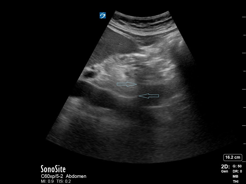
The following ultrasound is performed on a patient to evaluate their aorta.
1) What probe is being used? (2pts) 2) What axis of the body is this ultrasound in? (2pts) 3) What diameter aorta is considered "aneurysmic" (needs monitoring)? (2pts) 4) What is the name of the artifact responsible for displaying the arrowed discontinuity of the aorta? (4pts) (Courtesy of JG, JC) |
Question 2:
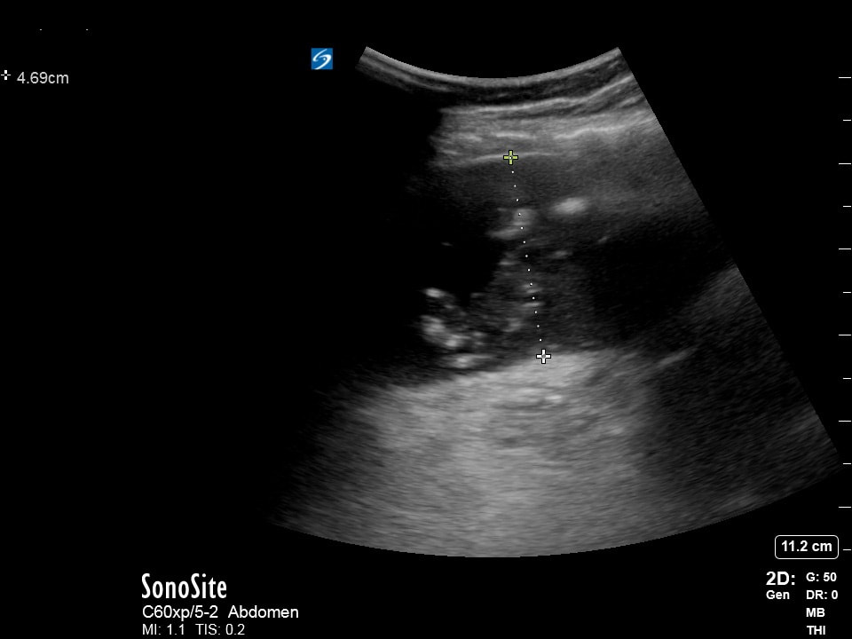
A middle aged patient presents after a MVC. The following ultrasound images are obtained and the patient is hemodynamically stable.
Clip A |
Clip B |
1) What exam is being performed? (2pts)
2) Is "Clip A" of the RUQ or LUQ? (2pts)
3) Is "Clip B" of the RUQ or LUQ? (2pts)
4) Assuming not other abnormalities found during the primary and secondary surveys, where does this patient need to go? (4pts)
(Courtesy of BM)
2) Is "Clip A" of the RUQ or LUQ? (2pts)
3) Is "Clip B" of the RUQ or LUQ? (2pts)
4) Assuming not other abnormalities found during the primary and secondary surveys, where does this patient need to go? (4pts)
(Courtesy of BM)
Question 3:
|
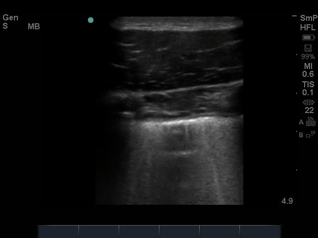
A patient presents with shortness of breath and the following ultrasound image is captured.
1) What probe is being used? (2pts) 2) What is being examined? (2pts) 3) How many b-lines per intercostal space suggest interstitial fluid? (2pts) 4) What causes b-lines/what kind of artifact create them? (4pts) (Courtesy of KG, AC, JH) |
Question 4:
|
The following cardiac ultrasound is performed.
1) What view is being attempted? (2 pts) 2) What is your interpretation? (2 pts) 3) What is most likely this patients chief complaint? (2 pts) 4) Name three physical exam findings this patient most likely has. (4 pts) (Courtesy of JC, JG) |
|
Question 5:
|
|
|
You are doing QA with your US director and see these images as part of a patient encounter.
1) What probe and mode are being used? (2pts)
2) What is being examined? (2pts)
3) What is most likely this patient's chief complaint? (2pts)
4) What is the most likely reason for this patient's chief complaint? (4pts)
(Courtesy of JC, JG)
1) What probe and mode are being used? (2pts)
2) What is being examined? (2pts)
3) What is most likely this patient's chief complaint? (2pts)
4) What is the most likely reason for this patient's chief complaint? (4pts)
(Courtesy of JC, JG)
Extra Credit:
|
Tyler... what the heck are you doing??? (Remember, points are given for correctness and creativity!!!)
1) What probe is being used? (2pts) 2) What is being examined? (2pts) 3) Which hospital was this ultrasound performed? (2pts) (Consider what machine was used to perform this ultrasound... or just guess... its a 50/50 shot!!!) 4) What does Tyler need to do next? (4pts) (Remember... points for creativity!!!) (Courtesy of TF) |
|