
Ultrasounds Of The Block
Welcome to SIU's EM Residency "Ultrasounds of the Block" curriculum (formerly known as "Ultrasound of the Month"). First instituted as a friendly competition to spark increased volume of bedside studies, it has now become a melee of ultrasound fun. As part of an asynchronous curriculum in emergency ultrasound, all ultrasounds performed at our clinical sites are reviewed for quality assurance by ultrasound director. Each block a few images/clips are selected because of an interesting finding, a high quality image showing great anatomy, or because they carry an important teaching point. These “Ultrasounds of the Block” are then displayed online with clinical vignettes and questions to test residents and their knowledge. The competition aspect of the curriculum is that residents gain points by performing any selected "Ultrasounds of the Block" and for answering monthly questions correctly. Each year a winner is announced based on overall points and surprised with trophy gifts to cherish and gloat about for the many years to come.
Past "Ultrasounds of the Block" can be viewed by clicking here. This blocks "Ultrasounds of the Block" can be viewed below.
Ultrasounds Of The Block (B10)
Question 1:
A patient presents with hypotension and the following ultrasound is performed as part of a RUSH exam. What type of shock are could the patient have and what is your next step in assessment? (10pts)
a) cardiogenic, obtain more views of the heart
b) cardiogenic, obtain more views of the lungs
c) obstructive, obtain more views of the heart
d) obstructive, obtain views of the lungs
Courtesy of SE
a) cardiogenic, obtain more views of the heart
b) cardiogenic, obtain more views of the lungs
c) obstructive, obtain more views of the heart
d) obstructive, obtain views of the lungs
Courtesy of SE
Question 2
The following ultrasound is performed on a patient with hypotension. What findings would be reassuring and suggest non-tamponade physiology on a SubX Long/IVC view in a spontaneously breathing patient? (10pts)
a) collapse > 50% of the IVC
b) dilation of the IVC
c) dilation of the hepatic veins
d) loss of respiratory variation of the IVC
e) b, c, & d
Courtesy of SE
a) collapse > 50% of the IVC
b) dilation of the IVC
c) dilation of the hepatic veins
d) loss of respiratory variation of the IVC
e) b, c, & d
Courtesy of SE
Question 3:
The following ultrasound is performed on a patient who arrested in route via EMS and achieved ROSC on your first pulse check? What was the most likely rhythm this patient presented with when the EMS started the code? (10pts)
a) asystole
b) PEA
c) vfib
d) vtach
Courtesy of SE
a) asystole
b) PEA
c) vfib
d) vtach
Courtesy of SE
Question 4:
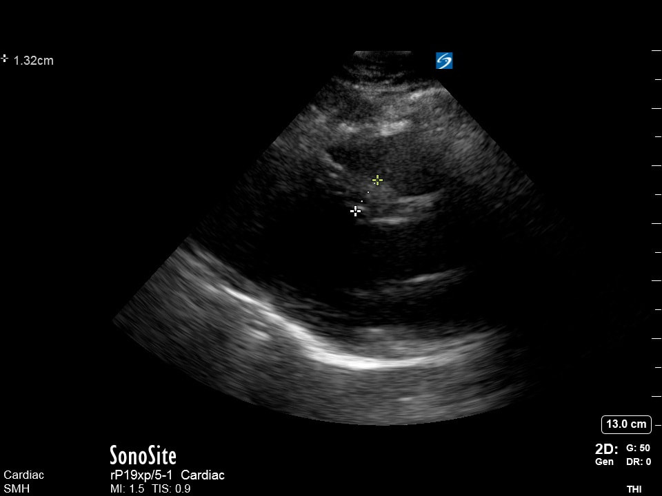
The following image is obtained at 3am at SMH on a 16 year old patient who presents with episodes of palpitations and dizziness for months. She has a family history of HOCM and a cardiology follow-up in 4 weeks, her vitals are stable and her PUF work-up including a CBC, CMP, dimer, EKG, and troponin (which was done 7 hours prior to you seeing her) was unremarkable. What would be the most appropriate next step in assessment and management? (10pts)
a) call pediatric cardiology for a consult
b) obtain a formal echocardiogram
c) obtain a pregnancy test (which was not ordered in PUF)
d) recommend to the patient and family they discuss with cardiology moving up their appointment, discharge them, and do a follow-up call the next day
Courtesy of AM
a) call pediatric cardiology for a consult
b) obtain a formal echocardiogram
c) obtain a pregnancy test (which was not ordered in PUF)
d) recommend to the patient and family they discuss with cardiology moving up their appointment, discharge them, and do a follow-up call the next day
Courtesy of AM