
Ultrasounds Of The Block
Welcome to SIU's EM Residency "Ultrasounds of the Block" curriculum (formerly known as "Ultrasound of the Month"). First instituted as a friendly competition to spark increased volume of bedside studies, it has now become a melee of ultrasound fun. As part of an asynchronous curriculum in emergency ultrasound, all ultrasounds performed at our clinical sites are reviewed for quality assurance by ultrasound director. Each block a few images/clips are selected because of an interesting finding, a high quality image showing great anatomy, or because they carry an important teaching point. These “Ultrasounds of the Block” are then displayed online with clinical vignettes and questions to test residents and their knowledge. The competition aspect of the curriculum is that residents gain points by performing any selected "Ultrasounds of the Block" and for answering monthly questions correctly. Each year a winner is announced based on overall points and surprised with trophy gifts to cherish and gloat about for the many years to come.
Past "Ultrasounds of the Block" can be viewed by clicking here. This blocks "Ultrasounds of the Block" can be viewed below.
Ultrasounds Of The Block (B3)
Question 1: The FAST and The furious
A 40 y/o male presents after an MVC where he was unrestrained. The following FAST is performed. Please answer the following for each image:
- The probe and exam setting being used (2pts)
- The view obtained (include axis and region/organ being examined) (2pts)
- List how the image could be optimized (3pts)
- Your interpretation of the image (3pts)
- The probe and exam setting being used (2pts)
- The view obtained (include axis and region/organ being examined) (2pts)
- List how the image could be optimized (3pts)
- Your interpretation of the image (3pts)
A)phased array, cardiac bmode
subX decrease depth beating, no effusion, no RVS, no LVD C)phased array, cardiac bmode
LUQ long axis switch to curvilinear & abdominal mode, decrease depth no free fluid |
B)phased array, cardiac bmode
RUQ long axis switch to curvilinear & abdominal mode free fluid D)phased array, cardiac bmode
bladder short axis switch to curvilinear & abdominal mode, decrease depth free fluid |
Extra Credit MCQ (5pts): An eFAST is completed on this same patient with the following thoracic windows obtained. The patients vitals are HR 120, BP 90/70, RR 30, sat of 80%, with an intact airway. Your staff has placed the patient on a monitor and have established 2 large bore IV’s. What is the next step in managing this patient?
(Left) |
(Right) |
(Choose the best answer)
A - Intravenous Crystalloids
B - Rapid Sequence Intubation (Airway is intact)
C - Thorocostomy (+ slide b/l on ultrasound)
D - Exploratory Laparotomy (definitive, but not the next best step from the choices, partial credit given for this)
A - Intravenous Crystalloids
B - Rapid Sequence Intubation (Airway is intact)
C - Thorocostomy (+ slide b/l on ultrasound)
D - Exploratory Laparotomy (definitive, but not the next best step from the choices, partial credit given for this)
(Courtesy of ET & NF)
Question 2: To FAST to Furious
A 40 y/o male presents after a hitting a stationary car while backing out of his parking spot. He was restrained and his only complaint was that he was feeling dizzy and lightheaded with palpitations prior to hitting the other car. The following FAST is performed. Please answer the following for each image:
- The probe and exam setting being used (2pts)
- The view obtained (include axis and region/organ being examined) (2pts)
- List how the image could be optimized (3pts)
- Your interpretation of the image (3pts)
- The probe and exam setting being used (2pts)
- The view obtained (include axis and region/organ being examined) (2pts)
- List how the image could be optimized (3pts)
- Your interpretation of the image (3pts)
A)phased array, cardiac bmode
SubX pretty good image pacer wires C)curvilinear, abdominal bmode
LUQ long axis decrease depth, roll on right side stomach in view, no free fluid |
B)curvilinear, abdominal bmode
RUQ long axis okay image no free fluid D)curvilinear, abdominal bmode
bladder short axis decrease depth no free fluid |
Extra Credit MCQ (5pts): During your evaluations the patient begins to start having palpitations, feels dizzy, passes out, and then has a full recovery. What is the next best step?
(Choose the best answer)
A - Intravenous Crystalloids
B - Pericardiocentesis
C - Emergent Cardiothoracic Surgery Consultation
D - Urgent Cardiology Consultation For Definitive Testing (Reccurrent syncope and has pacemaker, needs interrogation)
(Courtesy of ET & NF)
(Choose the best answer)
A - Intravenous Crystalloids
B - Pericardiocentesis
C - Emergent Cardiothoracic Surgery Consultation
D - Urgent Cardiology Consultation For Definitive Testing (Reccurrent syncope and has pacemaker, needs interrogation)
(Courtesy of ET & NF)
Question 3: Tokyo Drift
Two FAST exams are performed. Please provide your assessment on how these exams were performed including any corrective measures or techniques that need optimized if appropriate. FYI... You have to watch the whole video to see all the views. (10pts each)
A)cardiac - decent view
RUQ - abdominal mode and probe RUQ - do a LUQ view Bladder - abdominal mode and probe, decrease depth |
B)cardiac - fine if you are a cardiologist
RUQ - switch up your indicator, abdominal mode and probe LUQ - switch up your indicator, abdominal mode and probe Bladder - abdominal mode and probe, decrease depth |
(Courtesy of ET & NF)
Question 4: High Frequency... Image Delinquency...
|
|
The following ultrasound is obtained on a patient with difficulty breathing. Provide the following.
The probe and exam setting being used (2pts) The view obtained (include axis and region/organ being examined) (2pts) List how the image could be optimized (3pts) Your interpretation of the image (3pts) (Courtesy of MW) HFL, lung
long axis lung increase depth or switch to curvilinear (see added image) fluid in lung |
Question 5: The return of the Echo spot
The following echos were obtained. Please answer the following questions regarding each image.
|
|
A)What cardiac view is this? (2pt)
Is the heart beating? (2pt) Is there a pericardial effusion? (2pt) Is there signs of right heart strain? (2pt) Are there signs of left ventricular dysfunction? (2pt) PSL
Y N N N |
|
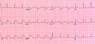
Extra Credit MCQ (5pts): If this echo was obtained in a facility > 2 hours away from a cath lab and the patient presented with the following EKG, what would be the next best choice in managing this patient?
(Choose the best answer) A - Aspirin B - Thrombolytics C - Labetolol (Look at the aortic root, it is almost 5cm, > 3.8cm worrisome for aneurysm or dissection) D - Atorvastatin |
|
|
B)What cardiac view is this? (2pt)
Is the heart beating? (2pt) Is there a pericardial effusion? (2pt) Is there signs of right heart strain? (2pt) Are there signs of left ventricular dysfunction? (2pt) SubX
Y Y N N Extra Credit MCQ (5pts): What EKG finding would you most likely see in this patient?
(Choose the best answer) A - Repolarization abnormalities in the precordial leads B - Delay in conduction between the P-wave and R-complex C - Variability in the voltage amplitude of the QRS complex (Electrical alternans) D - P-waves of varying morphology |
|
|
C)What cardiac view is this? (2pt)
Is the heart beating? (2pt) Is there a pericardial effusion? (2pt) Is there signs of right heart strain? (2pt) Are there signs of left ventricular dysfunction? (2pt) PSL
Y N N N |
|
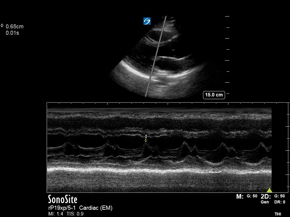
Extra Credit MCQ (5pts): The following image is obtained along with this ultrasound calculating the patients EPSS. At what distance is the EPSS highly suggestive of poor LV function?
(Choose the best answer) A - <7cm B - >7cm C - <7mm D - >7mm |
(Courtesy of JG, JH, & RT)