
Ultrasounds Of The Block
Welcome to SIU's EM Residency "Ultrasounds of the Block" curriculum (formerly known as "Ultrasound of the Month"). First instituted as a friendly competition to spark increased volume of bedside studies, it has now become a melee of ultrasound fun. As part of an asynchronous curriculum in emergency ultrasound, all ultrasounds performed at our clinical sites are reviewed for quality assurance by ultrasound director. Each block a few images/clips are selected because of an interesting finding, a high quality image showing great anatomy, or because they carry an important teaching point. These “Ultrasounds of the Block” are then displayed online with clinical vignettes and questions to test residents and their knowledge. The competition aspect of the curriculum is that residents gain points by performing any selected "Ultrasounds of the Block" and for answering monthly questions correctly. Each year a winner is announced based on overall points and surprised with trophy gifts to cherish and gloat about for the many years to come.
Past "Ultrasounds of the Block" can be viewed by clicking here. This blocks "Ultrasounds of the Block" can be viewed below.
Ultrasounds Of The Block (B4)
Question 1:The following ultrasound is performed on a patient with SOB
a) What probe and exam setting was used? (2pts) b) What is the most likely cause of this patients SOB? (2pts) c) What artifact helps you answer "b"? (2pts) d) What BLUE point is this probe most likely placed at? (i.e. upper right, lower right, upper left, lower left, PLAPS, etc) (4pts) Courtesy of ELI 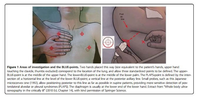
|
|
Question 2
|
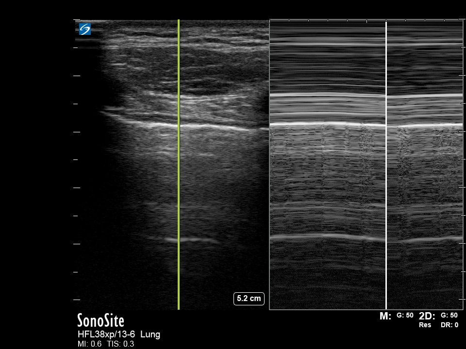
The following ultrasound is performed on a patient with SOB
a) What probe and exam setting was used? (2pts) b) What ultrasound mode is being used? (2pts) c) What is most likely causing this patients SOB? (2pts) d) What is the "gold standard" ultrasound finding that can confirm the suspected diagnosis? (4pts) Courtesy of CY & CP |
|
Question 3:
|
The following ultrasounds are performed as part of an assessment of a patient
a) What probe and exam setting was used? (2pts) b) What exam is most likely being peformed? (2pts) c) What view is missing? (2pts) d) What is your interpretation? (4pts) Courtesy of JL |
|
Question 4:
|
The following ultrasound is performed on a patient who is pale, diaphorectic, and clutching their chest
a) What probe and exam setting was used? (2pts) b) What view is being performed? (2pts) c) What is your interpretation? (4pts) d) What are your next steps in management? (2pts) Courtesy of AH, AM & ELI |
|